3EIF

1.9 angstrom crystal structure of the active form of the C5a peptidase from Streptococcus pyogenes (ScpA)


Experimental Data Snapshot

  • Method: X-RAY DIFFRACTION
  • Resolution: 1.90 Å
  • R-Value Free: 0.223 
  • R-Value Work: 0.191 
  • R-Value Observed: 0.191 

wwPDB Validation   3D Report Full Report


This is version 1.4 of the entry. See complete history


Literature

Model for Substrate Interactions in C5a Peptidase from Streptococcus pyogenes: A 1.9 A Crystal Structure of the Active Form of ScpA

Kagawa, T.F.O'Connell, M.R.Mouat, P.Paoli, M.O'Toole, P.W.Cooney, J.C.

(2009) J Mol Biol 386: 754-772

  • DOI: https://doi.org/10.1016/j.jmb.2008.12.074
  • Primary Citation of Related Structures:  
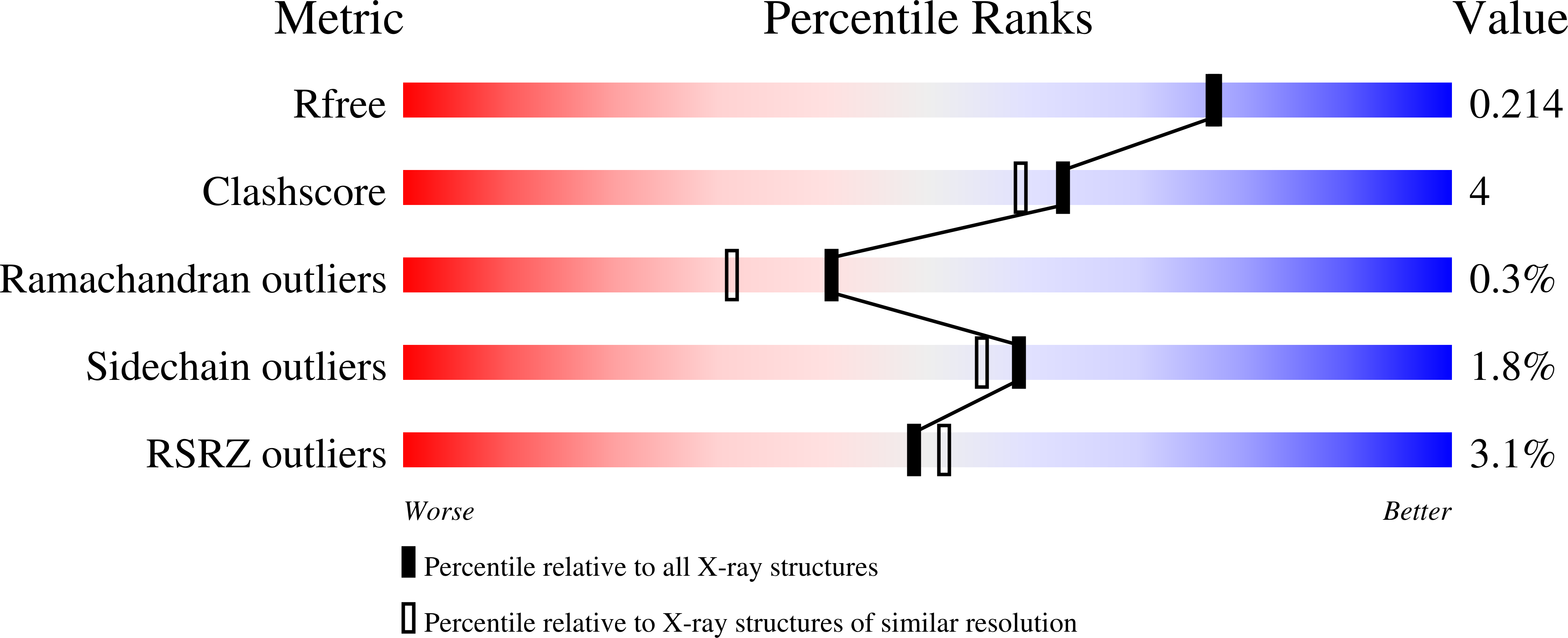
    3EIF

  • PubMed Abstract: 

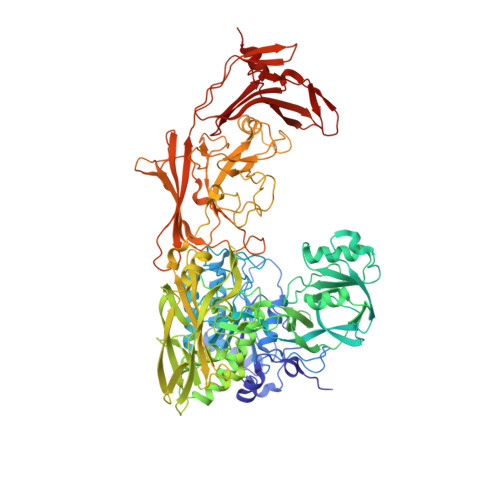
    The crystal structure of an active form of ScpA has been solved to 1.9 A resolution. ScpA is a multidomain cell-envelope subtilase from Streptococcus pyogenes that cleaves complement component C5a. The catalytic triad of ScpA is geometrically consistent with other subtilases, clearly demonstrating that the additional activation mechanism proposed for the Streptococcus agalactiae homologue (ScpB) is not required for ScpA. The ScpA structure revealed that access to the catalytic site is restricted by variable regions in the catalytic domain (vr7, vr9, and vr11) and by the presence of the inserted protease-associated (PA) domain and the second fibronectin type III domains (Fn2). Modeling of the ScpA-C5a complex indicates that the substrate binds with carboxyl-terminal residues (65-74) extended through the active site and core residues (1-64) forming exosite-type interactions with the Fn2 domain. This is reminiscent of the two-site mechanism proposed for C5a binding to its receptor. In the nonprime region of the active site, interactions with the substrate backbone are predicted to be more similar to those observed in kexins, involving a single beta-strand in the peptidase. However, in contrast to kexins, there would be diminished emphasis on side-chain interactions, with little charged character in the S3-S1 and S6-S4 subsites occupied by the side chains of residues in vr7 and vr9. Substrate binding is anticipated to be dominated by ionic interactions in two distinct regions of ScpA. On the prime side of the active site, salt bridges are predicted between P1', P2', and P7' residues, and residues in the catalytic and PA domains. Remote to the active site, a larger number of ionic interactions between residues in the C5a core and the Fn2 domain are observed in the model. Thus, both PA and Fn2 domains are expected to play significant roles in substrate recognition.


  • Organizational Affiliation

    Department of Life Sciences, University of Limerick, Limerick, Ireland. kagawatf@gmail.com


Macromolecules
Find similar proteins by:  (by identity cutoff)  |  3D Structure
Entity ID: 1
MoleculeChains Sequence LengthOrganismDetailsImage
C5a peptidase936Streptococcus pyogenesMutation(s): 1 
Gene Names: scpA
EC: 3.4.21.110
UniProt
Find proteins for B6ETQ5 (Streptococcus pyogenes)
Explore B6ETQ5 
Go to UniProtKB:  B6ETQ5
Entity Groups  
Sequence Clusters30% Identity50% Identity70% Identity90% Identity95% Identity100% Identity
UniProt GroupB6ETQ5
Sequence Annotations
Expand
  • Reference Sequence
Small Molecules
Ligands 5 Unique
IDChains Name / Formula / InChI Key2D Diagram3D Interactions
EPE
Query on EPE

Download Ideal Coordinates CCD File 
K [auth A]4-(2-HYDROXYETHYL)-1-PIPERAZINE ETHANESULFONIC ACID
C8 H18 N2 O4 S
JKMHFZQWWAIEOD-UHFFFAOYSA-N
MLA
Query on MLA

Download Ideal Coordinates CCD File 
L [auth A]MALONIC ACID
C3 H4 O4
OFOBLEOULBTSOW-UHFFFAOYSA-N
SO4
Query on SO4

Download Ideal Coordinates CCD File 
D [auth A]
E [auth A]
F [auth A]
G [auth A]
H [auth A]
D [auth A],
E [auth A],
F [auth A],
G [auth A],
H [auth A],
I [auth A],
J [auth A]
SULFATE ION
O4 S
QAOWNCQODCNURD-UHFFFAOYSA-L
CA
Query on CA

Download Ideal Coordinates CCD File 
B [auth A]CALCIUM ION
Ca
BHPQYMZQTOCNFJ-UHFFFAOYSA-N
NA
Query on NA

Download Ideal Coordinates CCD File 
C [auth A]SODIUM ION
Na
FKNQFGJONOIPTF-UHFFFAOYSA-N
Experimental Data & Validation

Experimental Data

  • Method: X-RAY DIFFRACTION
  • Resolution: 1.90 Å
  • R-Value Free: 0.223 
  • R-Value Work: 0.191 
  • R-Value Observed: 0.191 
  • Space Group: P 63 2 2
Unit Cell:
Length ( Å )Angle ( ˚ )
a = 167.561α = 90
b = 167.561β = 90
c = 141.759γ = 120
Software Package:
Software NamePurpose
ADSCdata collection
PHASERphasing
CNSrefinement
MOSFLMdata reduction
SCALAdata scaling

Structure Validation

View Full Validation Report


Entry History 

Deposition Data

Revision History  (Full details and data files)

  • Version 1.0: 2009-02-24
    Type: Initial release
  • Version 1.1: 2011-07-13
    Changes: Version format compliance
  • Version 1.2: 2015-04-29
    Changes: Non-polymer description
  • Version 1.3: 2021-11-10
    Changes: Database references, Derived calculations
  • Version 1.4: 2023-11-01
    Changes: Data collection, Refinement description