


X-ray crystal structure of N-acetylglucosamine-6-phosphate deacetylase from Vibrio cholerae.
Osipiuk, J., Maltseva, N., Stam, J., Anderson, W.F., Joachimiak, A.To be published.
Experimental Data Snapshot
wwPDB Validation 3D Report Full Report

Entity ID: 1 | |||||
|---|---|---|---|---|---|
| Molecule | Chains | Sequence Length | Organism | Details | Image |
| N-acetylglucosamine-6-phosphate deacetylase | 381 | Vibrio cholerae | Mutation(s): 0 Gene Names: nagA, VC_0994 EC: 3.5.1.25 | 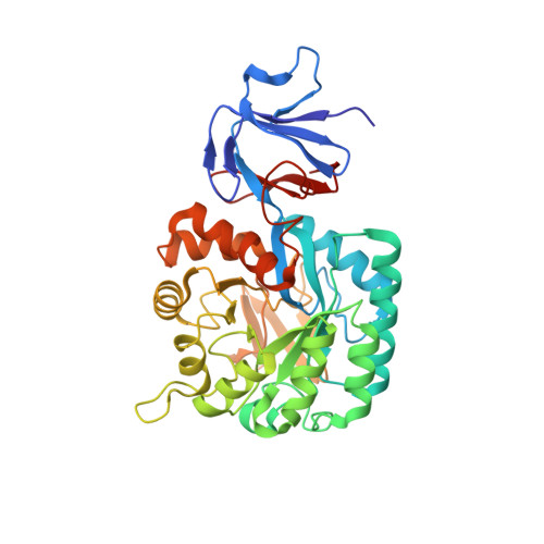 | |
UniProt | |||||
Find proteins for O32445 (Vibrio cholerae serotype O1 (strain ATCC 39315 / El Tor Inaba N16961)) Explore O32445 Go to UniProtKB: O32445 | |||||
Entity Groups | |||||
| Sequence Clusters | 30% Identity50% Identity70% Identity90% Identity95% Identity100% Identity | ||||
| UniProt Group | O32445 | ||||
Sequence AnnotationsExpand | |||||
| |||||
| Ligands 2 Unique | |||||
|---|---|---|---|---|---|
| ID | Chains | Name / Formula / InChI Key | 2D Diagram | 3D Interactions | |
| SO4 Query on SO4 | D [auth A], F [auth B] | SULFATE ION O4 S QAOWNCQODCNURD-UHFFFAOYSA-L |  | ||
| NI Query on NI | C [auth A], E [auth B] | NICKEL (II) ION Ni VEQPNABPJHWNSG-UHFFFAOYSA-N |  | ||
| Length ( Å ) | Angle ( ˚ ) |
|---|---|
| a = 77.001 | α = 90 |
| b = 77.001 | β = 90 |
| c = 282.549 | γ = 90 |
| Software Name | Purpose |
|---|---|
| DENZO | data reduction |
| SCALEPACK | data scaling |
| REFMAC | refinement |
| PDB_EXTRACT | data extraction |
| SBC-Collect | data collection |
| HKL-3000 | data reduction |
| MOLREP | phasing |
RCSB PDB (citation) is hosted by
RCSB PDB is a member of the

