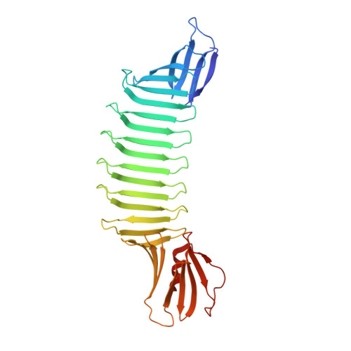
Molecular mechanism of thioflavin-T binding to the surface of beta-rich peptide self-assemblies
Biancalana, M., Makabe, K., Koide, A., Koide, S.(2009) J Mol Biol 385: 1052-1063
- PubMed: 19038267
- DOI: https://doi.org/10.1016/j.jmb.2008.11.006
- Primary Citation of Related Structures:
3EC5 - PubMed Abstract:
A number of small organic molecules have been developed that bind to amyloid fibrils, a subset of which also inhibit fibrillization. Among these, the benzothiol dye Thioflavin-T (ThT) has been used for decades in the diagnosis of protein-misfolding diseases and in kinetic studies of self-assembly (fibrillization). Despite its importance, efforts to characterize the ThT-binding mechanism at the atomic level have been hampered by the inherent insolubility and heterogeneity of peptide self-assemblies. To overcome these challenges, we have developed a minimalist approach to designing a ThT-binding site in a "peptide self-assembly mimic" (PSAM) scaffold. PSAMs are engineered water-soluble proteins that mimic a segment of beta-rich peptide self-assembly, and they are amenable to standard biophysical techniques and systematic mutagenesis. The PSAM beta-sheet contains rows of repetitive amino acid patterns running perpendicular to the strands (cross-strand ladders) that represent a ubiquitous structural feature of fibril-like surfaces. We successfully designed a ThT-binding site that recapitulates the hallmarks of ThT-fibril interactions by constructing a cross-strand ladder consisting of contiguous tyrosines. The X-ray crystal structures suggest that ThT interacts with the beta-sheet by docking onto surfaces formed by a single tyrosine ladder, rather than in the space between adjacent ladders. Systematic mutagenesis further demonstrated that tyrosine surfaces across four or more beta-strands formed the minimal binding site for ThT. Our work thus provides structural insights into how this widely used dye recognizes a prominent subset of peptide self-assemblies, and proposes a strategy to elucidate the mechanisms of fibril-ligand interactions.
Organizational Affiliation:
Department of Biochemistry and Molecular Biology, The University of Chicago, 929 East 57th Street, Chicago, IL 60637, USA.