3E80

Structure of Heparinase II complexed with heparan sulfate degradation disaccharide product


Experimental Data Snapshot

  • Method: X-RAY DIFFRACTION
  • Resolution: 2.35 Å
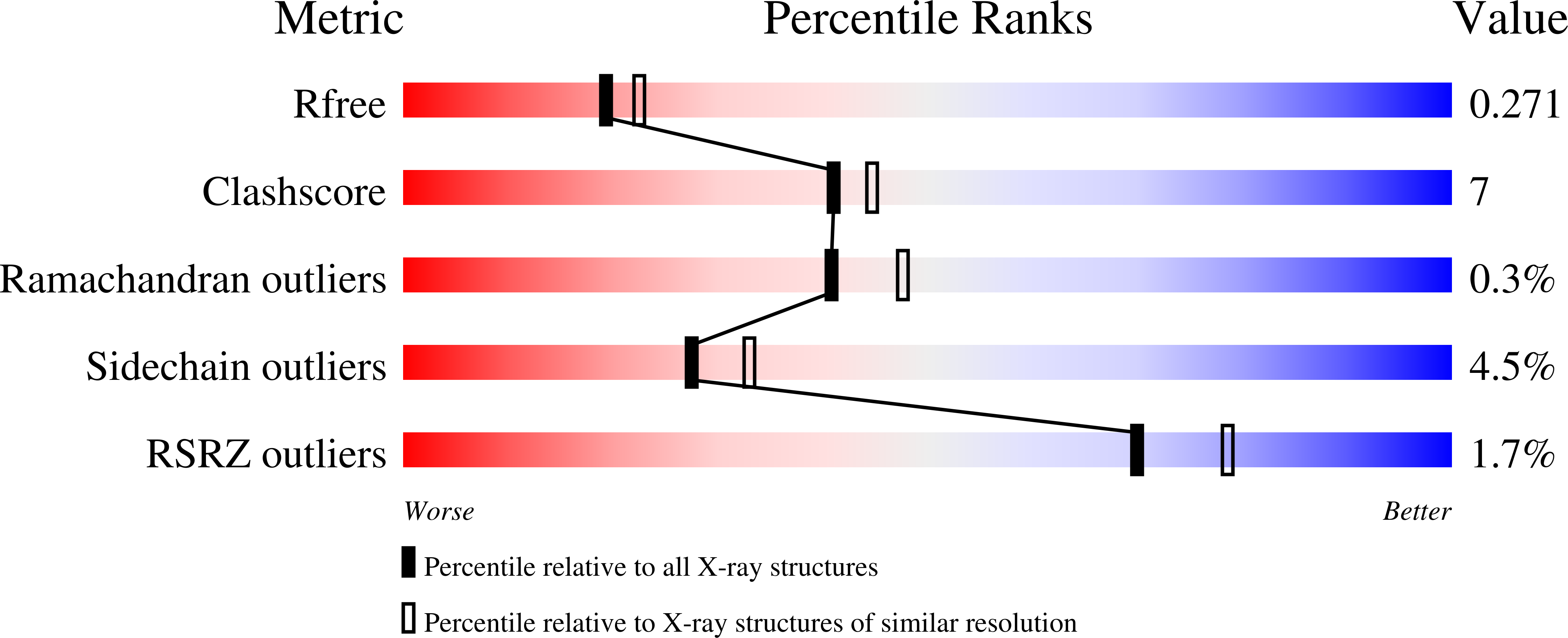
  • R-Value Free: 0.267 
  • R-Value Work: 0.229 
  • R-Value Observed: 0.231 

wwPDB Validation   3D Report Full Report


This is version 3.1 of the entry. See complete history


Literature

Catalytic mechanism of heparinase II investigated by site-directed mutagenesis and the crystal structure with its substrate.

Shaya, D.Zhao, W.Garron, M.L.Xiao, Z.Cui, Q.Zhang, Z.Sulea, T.Linhardt, R.J.Cygler, M.

(2010) J Biol Chem 285: 20051-20061

  • DOI: https://doi.org/10.1074/jbc.M110.101071
  • Primary Citation of Related Structures:  
    3E7J, 3E80

  • PubMed Abstract: 

    Heparinase II (HepII) is an 85-kDa dimeric enzyme that depolymerizes both heparin and heparan sulfate glycosaminoglycans through a beta-elimination mechanism. Recently, we determined the crystal structure of HepII from Pedobacter heparinus (previously known as Flavobacterium heparinum) in complex with a heparin disaccharide product, and identified the location of its active site. Here we present the structure of HepII complexed with a heparan sulfate disaccharide product, proving that the same binding/active site is responsible for the degradation of both uronic acid epimers containing substrates. The key enzymatic step involves removal of a proton from the C5 carbon (a chiral center) of the uronic acid, posing a topological challenge to abstract the proton from either side of the ring in a single active site. We have identified three potential active site residues equidistant from C5 and located on both sides of the uronate product and determined their role in catalysis using a set of defined tetrasaccharide substrates. HepII H202A/Y257A mutant lost activity for both substrates and we determined its crystal structure complexed with a heparan sulfate-derived tetrasaccharide. Based on kinetic characterization of various mutants and the structure of the enzyme-substrate complex we propose residues participating in catalysis and their specific roles.


  • Organizational Affiliation

    Department of Biochemistry, McGill University, Montréal, Québec H3G 1Y6, Canada.


Macromolecules
Find similar proteins by:  (by identity cutoff)  |  3D Structure
Entity ID: 1
MoleculeChains Sequence LengthOrganismDetailsImage
Heparinase II protein
A, B, C
749Pedobacter heparinusMutation(s): 0 
UniProt
Find proteins for C6XZB6 (Pedobacter heparinus (strain ATCC 13125 / DSM 2366 / CIP 104194 / JCM 7457 / NBRC 12017 / NCIMB 9290 / NRRL B-14731 / HIM 762-3))
Explore C6XZB6 
Go to UniProtKB:  C6XZB6
Entity Groups  
Sequence Clusters30% Identity50% Identity70% Identity90% Identity95% Identity100% Identity
UniProt GroupC6XZB6
Sequence Annotations
Expand
  • Reference Sequence
Oligosaccharides

Help

Entity ID: 2
MoleculeChains Length2D Diagram Glycosylation3D Interactions
alpha-D-xylopyranose-(1-4)-alpha-D-glucopyranuronic acid-(1-2)-[alpha-L-rhamnopyranose-(1-4)]alpha-D-mannopyranose
D, E, F
4O-Glycosylation
Glycosylation Resources
GlyTouCan:  G51375MF
GlyCosmos:  G51375MF
Entity ID: 3
MoleculeChains Length2D Diagram Glycosylation3D Interactions
4-deoxy-alpha-L-threo-hex-4-enopyranuronic acid-(1-4)-2-acetamido-2-deoxy-alpha-D-glucopyranose
G, H, I
2N/A
Glycosylation Resources
GlyTouCan:  G54188YF
GlyCosmos:  G54188YF
Small Molecules
Ligands 2 Unique
IDChains Name / Formula / InChI Key2D Diagram3D Interactions
PO4
Query on PO4

Download Ideal Coordinates CCD File 
K [auth A]
L [auth A]
M [auth A]
N [auth A]
O [auth B]
K [auth A],
L [auth A],
M [auth A],
N [auth A],
O [auth B],
Q [auth B],
R [auth B],
T [auth C],
U [auth C]
PHOSPHATE ION
O4 P
NBIIXXVUZAFLBC-UHFFFAOYSA-K
ZN
Query on ZN

Download Ideal Coordinates CCD File 
J [auth A],
P [auth B],
S [auth C]
ZINC ION
Zn
PTFCDOFLOPIGGS-UHFFFAOYSA-N
Modified Residues  1 Unique
IDChains TypeFormula2D DiagramParent
PCA
Query on PCA
A, B, C
L-PEPTIDE LINKINGC5 H7 N O3GLN
Experimental Data & Validation

Experimental Data

  • Method: X-RAY DIFFRACTION
  • Resolution: 2.35 Å
  • R-Value Free: 0.267 
  • R-Value Work: 0.229 
  • R-Value Observed: 0.231 
  • Space Group: P 21 21 2
Unit Cell:
Length ( Å )Angle ( ˚ )
a = 201.28α = 90
b = 209.36β = 90
c = 59.22γ = 90
Software Package:
Software NamePurpose
d*TREKdata scaling
REFMACrefinement
PDB_EXTRACTdata extraction
d*TREKdata reduction
MOLREPphasing

Structure Validation

View Full Validation Report


Entry History 

Deposition Data

Revision History  (Full details and data files)

  • Version 1.0: 2008-12-30
    Type: Initial release
  • Version 1.1: 2011-07-13
    Changes: Advisory, Version format compliance
  • Version 2.0: 2019-12-25
    Changes: Advisory, Data collection, Database references, Derived calculations, Polymer sequence
  • Version 3.0: 2020-07-29
    Type: Remediation
    Reason: Carbohydrate remediation
    Changes: Advisory, Atomic model, Data collection, Derived calculations, Non-polymer description, Structure summary
  • Version 3.1: 2023-08-30
    Changes: Data collection, Database references, Refinement description, Structure summary