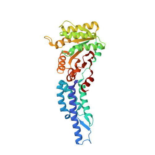
3DMD

Structures and Conformations in Solution of the Signal Recognition Particle Receptor from the archaeon Pyrococcus furiosus


Experimental Data Snapshot

  • Method: X-RAY DIFFRACTION
  • Resolution: 2.21 Å
  • R-Value Free: 0.242 
  • R-Value Work: 0.189 
  • R-Value Observed: 0.191 

wwPDB Validation   3D Report Full Report


This is version 1.3 of the entry. See complete history


Literature

Structures of the Signal Recognition Particle Receptor from the Archaeon Pyrococcus furiosus: Implications for the Targeting Step at the Membrane.

Egea, P.F.Tsuruta, H.de Leon, G.P.Napetschnig, J.Walter, P.Stroud, R.M.

(2008) PLoS One 3: e3619-e3619

  • DOI: https://doi.org/10.1371/journal.pone.0003619
  • Primary Citation of Related Structures:  
    3DM9, 3DMD, 3E70

  • PubMed Abstract: 

    In all organisms, a ribonucleoprotein called the signal recognition particle (SRP) and its receptor (SR) target nascent proteins from the ribosome to the translocon for secretion or membrane insertion. We present the first X-ray structures of an archeal FtsY, the receptor from the hyper-thermophile Pyrococcus furiosus (Pfu), in its free and GDP*magnesium-bound forms. The highly charged N-terminal domain of Pfu-FtsY is distinguished by a long N-terminal helix. The basic charges on the surface of this helix are likely to regulate interactions at the membrane. A peripheral GDP bound near a regulatory motif could indicate a site of interaction between the receptor and ribosomal or SRP RNAs. Small angle X-ray scattering and analytical ultracentrifugation indicate that the crystal structure of Pfu-FtsY correlates well with the average conformation in solution. Based on previous structures of two sub-complexes, we propose a model of the core of archeal and eukaryotic SRP*SR targeting complexes.


  • Organizational Affiliation

    Department of Biochemistry and Biophysics, University of California San Francisco, San Francisco, California, USA. pascal@msg.ucsf.edu


Macromolecules
Find similar proteins by:  (by identity cutoff)  |  3D Structure
Entity ID: 1
MoleculeChains Sequence LengthOrganismDetailsImage
Signal recognition particle receptorA [auth B],
B [auth A],
C
328Pyrococcus furiosusMutation(s): 0 
Gene Names: PF1766
UniProt
Find proteins for Q8U051 (Pyrococcus furiosus (strain ATCC 43587 / DSM 3638 / JCM 8422 / Vc1))
Explore Q8U051 
Go to UniProtKB:  Q8U051
Entity Groups  
Sequence Clusters30% Identity50% Identity70% Identity90% Identity95% Identity100% Identity
UniProt GroupQ8U051
Sequence Annotations
Expand
  • Reference Sequence
Small Molecules
Ligands 2 Unique
IDChains Name / Formula / InChI Key2D Diagram3D Interactions
SO4
Query on SO4

Download Ideal Coordinates CCD File 
D [auth B]
E [auth B]
F [auth B]
H [auth A]
I [auth A]
D [auth B],
E [auth B],
F [auth B],
H [auth A],
I [auth A],
J [auth A],
K [auth A],
M [auth C],
N [auth C],
O [auth C]
SULFATE ION
O4 S
QAOWNCQODCNURD-UHFFFAOYSA-L
GOL
Query on GOL

Download Ideal Coordinates CCD File 
G [auth B]
L [auth A]
P [auth C]
Q [auth C]
R [auth C]
G [auth B],
L [auth A],
P [auth C],
Q [auth C],
R [auth C],
S [auth C]
GLYCEROL
C3 H8 O3
PEDCQBHIVMGVHV-UHFFFAOYSA-N
Experimental Data & Validation

Experimental Data

  • Method: X-RAY DIFFRACTION
  • Resolution: 2.21 Å
  • R-Value Free: 0.242 
  • R-Value Work: 0.189 
  • R-Value Observed: 0.191 
  • Space Group: C 1 2 1
Unit Cell:
Length ( Å )Angle ( ˚ )
a = 149.956α = 90
b = 102.368β = 119.81
c = 101.766γ = 90
Software Package:
Software NamePurpose
PHENIXrefinement
ELVESrefinement
MOSFLMdata reduction
SCALAdata scaling
PHENIXphasing

Structure Validation

View Full Validation Report


Entry History 

Deposition Data

Revision History  (Full details and data files)

  • Version 1.0: 2008-11-11
    Type: Initial release
  • Version 1.1: 2011-07-13
    Changes: Non-polymer description, Version format compliance
  • Version 1.2: 2017-10-25
    Changes: Refinement description
  • Version 1.3: 2023-08-30
    Changes: Data collection, Database references, Derived calculations, Refinement description