Changing the determinants of protein stability from covalent to non-covalent interactions by in vitro evolution: a structural and energetic analysis.
Kather, I., Jakob, R.P., Dobbek, H., Schmid, F.X.(2008) J Mol Biol 381: 1040-1054
- PubMed: 18621056
- DOI: https://doi.org/10.1016/j.jmb.2008.06.073
- Primary Citation of Related Structures:
3DGS - PubMed Abstract:
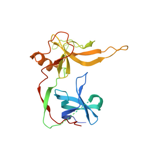
The three disulfide bonds of the gene-3-protein of the phage fd are essential for the conformational stability of this protein, and it unfolds when they are removed by reduction or mutation. Previously, we used an iterative in vitro selection strategy to generate a stable and functional form of the gene-3-protein without these disulfides. It yielded optimal replacements for the disulfide bonds as well as several stabilizing second-site mutations. The best selected variant showed a higher thermal stability compared with the disulfide-bonded wild-type protein. Here, we investigated the molecular basis of this strong stabilization by solving the crystal structure of this variant and by analyzing the contributions to the conformational stability of the selected mutations individually. They could mostly be explained by improved side-chain packing. The R29W substitution alone increased the midpoint of the thermal unfolding transition by 14 deg and the conformational stability by about 25 kJ mol(-1). This key mutation (i) removed a charged side chain that forms a buried salt bridge in the disulfide-containing wild-type protein, (ii) optimized the local packing with the residues that replace the C46-C53 disulfide and (iii) improved the domain interactions. Apparently, certain residues in proteins indeed play key roles for stability.
Organizational Affiliation:
Laboratorium für Biochemie, Universitat Bayreuth, 95440 Bayreuth, Germany.


