


Structural and Enzymatic Characterization of a Thermostable PQQ-dependent Soluble Aldose Sugar Dehydrogenase
Southall, S.M., Doel, J.J., Oubrie, A., Richardson, D.J.To be published.
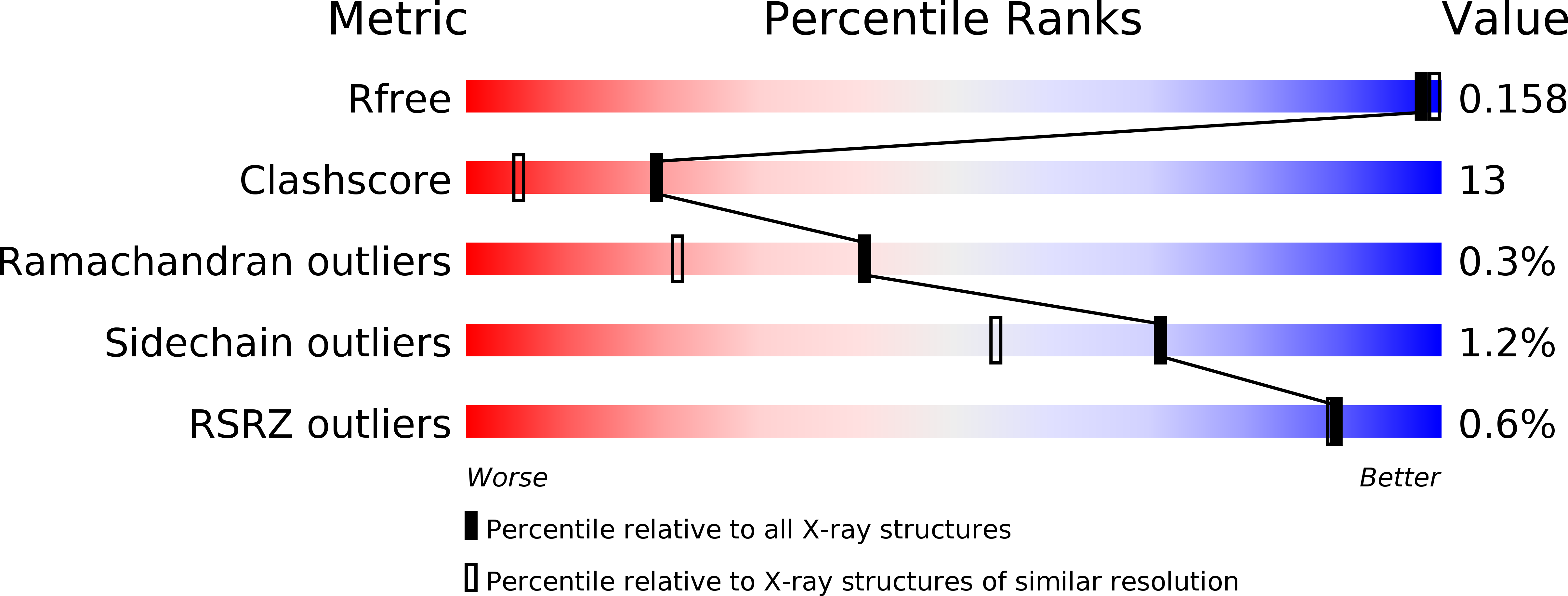
Experimental Data Snapshot
Entity ID: 1 | |||||
|---|---|---|---|---|---|
| Molecule | Chains | Sequence Length | Organism | Details | Image |
| Putative oxidoreductase | 347 | Streptomyces coelicolor | Mutation(s): 0 | 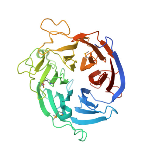 | |
UniProt | |||||
Find proteins for Q9Z571 (Streptomyces coelicolor (strain ATCC BAA-471 / A3(2) / M145)) Explore Q9Z571 Go to UniProtKB: Q9Z571 | |||||
Entity Groups | |||||
| Sequence Clusters | 30% Identity50% Identity70% Identity90% Identity95% Identity100% Identity | ||||
| UniProt Group | Q9Z571 | ||||
Sequence AnnotationsExpand | |||||
| |||||
| Ligands 4 Unique | |||||
|---|---|---|---|---|---|
| ID | Chains | Name / Formula / InChI Key | 2D Diagram | 3D Interactions | |
| PQQ Query on PQQ | F [auth A] | PYRROLOQUINOLINE QUINONE C14 H6 N2 O8 MMXZSJMASHPLLR-UHFFFAOYSA-N |  | ||
| ARA Query on ARA | B [auth A], C [auth A] | alpha-L-arabinopyranose C5 H10 O5 SRBFZHDQGSBBOR-QMKXCQHVSA-N |  | ||
| EDO Query on EDO | G [auth A] H [auth A] I [auth A] J [auth A] K [auth A] | 1,2-ETHANEDIOL C2 H6 O2 LYCAIKOWRPUZTN-UHFFFAOYSA-N |  | ||
| CA Query on CA | D [auth A], E [auth A] | CALCIUM ION Ca BHPQYMZQTOCNFJ-UHFFFAOYSA-N |  | ||
| Modified Residues 1 Unique | |||||
|---|---|---|---|---|---|
| ID | Chains | Type | Formula | 2D Diagram | Parent |
| MSE Query on MSE | A | L-PEPTIDE LINKING | C5 H11 N O2 Se |  | MET |
| Length ( Å ) | Angle ( ˚ ) |
|---|---|
| a = 49.033 | α = 90 |
| b = 41.26 | β = 93.58 |
| c = 85.931 | γ = 90 |
| Software Name | Purpose |
|---|---|
| REFMAC | refinement |
| MAR345dtb | data collection |
| MOSFLM | data reduction |
| SCALA | data scaling |
| MLPHARE | phasing |
RCSB PDB (citation) is hosted by
RCSB PDB is a member of the

