3CGA

Crystal structure of metastasis-associated protein S100A4 in the active, calcium-bound form


Experimental Data Snapshot

  • Method: X-RAY DIFFRACTION
  • Resolution: 2.03 Å
  • R-Value Free: 0.331 
  • R-Value Observed: 0.255 

wwPDB Validation   3D Report Full Report


This is version 1.2 of the entry. See complete history


Literature

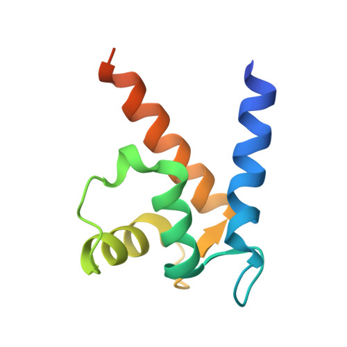
Crystal structure of metastasis-associated protein S100A4 in the active calcium-bound form

Pathuri, P.Vogeley, L.Luecke, H.

(2008) J Mol Biol 383: 62-77

  • DOI: https://doi.org/10.1016/j.jmb.2008.04.076
  • Primary Citation of Related Structures:  
    3CGA

  • PubMed Abstract: 

    S100A4 (metastasin) is a member of the S100 family of calcium-binding proteins that is directly involved in tumorigenesis. Until recently, the only structural information available was the solution NMR structure of the inactive calcium-free form of the protein. Here we report the crystal structure of human S100A4 in the active calcium-bound state at 2.03 A resolution that was solved by molecular replacement in the space group P6(5) with two molecules in the asymmetric unit from perfectly merohedrally twinned crystals. The Ca(2+)-bound S100A4 structure reveals a large conformational change in the three-dimensional structure of the dimeric S100A4 protein upon calcium binding. This calcium-dependent conformational change opens up a hydrophobic binding pocket that is capable of binding to target proteins such as annexin A2, the tumor-suppressor protein p53 and myosin IIA. The structure of the active form of S100A4 provides insight into its interactions with its binding partners and a better understanding of its role in metastasis.


  • Organizational Affiliation

    Department of Molecular Biology and Biochemistry, University of California, Irvine, CA 92697, USA.


Macromolecules
Find similar proteins by:  (by identity cutoff)  |  3D Structure
Entity ID: 1
MoleculeChains Sequence LengthOrganismDetailsImage
Protein S100-A4
A, B
101Homo sapiensMutation(s): 0 
Gene Names: S100A4CAPLMTS1
UniProt & NIH Common Fund Data Resources
Find proteins for P26447 (Homo sapiens)
Explore P26447 
Go to UniProtKB:  P26447
PHAROS:  P26447
GTEx:  ENSG00000196154 
Entity Groups  
Sequence Clusters30% Identity50% Identity70% Identity90% Identity95% Identity100% Identity
UniProt GroupP26447
Sequence Annotations
Expand
  • Reference Sequence
Experimental Data & Validation

Experimental Data

  • Method: X-RAY DIFFRACTION
  • Resolution: 2.03 Å
  • R-Value Free: 0.331 
  • R-Value Observed: 0.255 
  • Space Group: P 65
Unit Cell:
Length ( Å )Angle ( ˚ )
a = 47.107α = 90
b = 47.107β = 90
c = 176.2γ = 120
Software Package:
Software NamePurpose
SHELXmodel building
SHELXL-97refinement
DENZOdata reduction
HKL-2000data scaling
PHASERphasing

Structure Validation

View Full Validation Report


Entry History 

Deposition Data

Revision History  (Full details and data files)

  • Version 1.0: 2008-09-30
    Type: Initial release
  • Version 1.1: 2011-07-13
    Changes: Version format compliance
  • Version 1.2: 2024-02-21
    Changes: Data collection, Database references, Derived calculations