3C9I

Structure of P22 Tail-Needle GP26 Bound to Xenon Gas


Experimental Data Snapshot

  • Method: X-RAY DIFFRACTION
  • Resolution: 1.95 Å
  • R-Value Free: 0.269 
  • R-Value Work: 0.234 
  • R-Value Observed: 0.234 

wwPDB Validation   3D Report Full Report


This is version 1.3 of the entry. See complete history


Literature

Structural plasticity of the phage P22 tail needle gp26 probed with xenon gas.

Olia, A.S.Casjens, S.Cingolani, G.

(2009) Protein Sci 18: 537-548

  • DOI: https://doi.org/10.1002/pro.53
  • Primary Citation of Related Structures:  
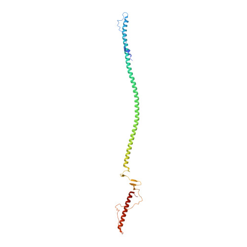
    3C9I

  • PubMed Abstract: 

    The tail needle, gp26, is a highly stable homo-trimeric fiber found in the tail apparatus of bacteriophage P22. In the mature virion, gp26 is responsible for plugging the DNA exit channel, and likely plays an important role in penetrating the host cell envelope. In this article, we have determined the 1.98 A resolution crystal structure of gp26 bound to xenon gas. The structure led us to identify a calcium and a chloride ion intimately bound at the interior of alpha-helical core, as well as seven small cavities occupied by xenon atoms. The two ions engage in buried polar interactions with gp26 side chains that provide specificity and register to gp26 helical core, thus enhancing its stability. Conversely, the distribution of xenon accessible cavities correlates well with the flexibility of the fiber observed in solution and in the crystal structure. We suggest that small internal cavities in gp26 between the helical core and the C-terminal tip allow for flexible swinging of the latter, without affecting the overall stability of the protein. The C-terminal tip may be important in scanning the bacterial surface in search of a cell-envelope penetration site, or for recognition of a yet unidentified receptor on the surface of the host.


  • Organizational Affiliation

    Department of Biochemistry and Molecular Biology, SUNY Upstate Medical University, Syracuse, New York 13210, USA.


Macromolecules
Find similar proteins by:  (by identity cutoff)  |  3D Structure
Entity ID: 1
MoleculeChains Sequence LengthOrganismDetailsImage
Tail needle protein gp26
A, B, C, D, E
A, B, C, D, E, F
242N/AMutation(s): 2 
Gene Names: 26
UniProt
Find proteins for P35837 (Salmonella phage P22)
Explore P35837 
Go to UniProtKB:  P35837
Entity Groups  
Sequence Clusters30% Identity50% Identity70% Identity90% Identity95% Identity100% Identity
UniProt GroupP35837
Sequence Annotations
Expand
  • Reference Sequence
Small Molecules
Ligands 3 Unique
IDChains Name / Formula / InChI Key2D Diagram3D Interactions
XE
Query on XE

Download Ideal Coordinates CCD File 
G [auth A]
H [auth A]
I [auth A]
J [auth A]
K [auth A]
G [auth A],
H [auth A],
I [auth A],
J [auth A],
K [auth A],
L [auth A],
O [auth C],
P [auth D],
Q [auth D],
R [auth D],
S [auth D],
T [auth D],
U [auth D],
X [auth F]
XENON
Xe
FHNFHKCVQCLJFQ-UHFFFAOYSA-N
CA
Query on CA

Download Ideal Coordinates CCD File 
M [auth A],
V [auth D]
CALCIUM ION
Ca
BHPQYMZQTOCNFJ-UHFFFAOYSA-N
CL
Query on CL

Download Ideal Coordinates CCD File 
N [auth A],
W [auth D]
CHLORIDE ION
Cl
VEXZGXHMUGYJMC-UHFFFAOYSA-M
Experimental Data & Validation

Experimental Data

  • Method: X-RAY DIFFRACTION
  • Resolution: 1.95 Å
  • R-Value Free: 0.269 
  • R-Value Work: 0.234 
  • R-Value Observed: 0.234 
  • Space Group: P 1 21 1
Unit Cell:
Length ( Å )Angle ( ˚ )
a = 40.539α = 90
b = 114.531β = 91.01
c = 172.045γ = 90
Software Package:
Software NamePurpose
ADSCdata collection
MOLREPphasing
CNSrefinement
HKL-2000data reduction
HKL-2000data scaling

Structure Validation

View Full Validation Report


Entry History 

Deposition Data

Revision History  (Full details and data files)

  • Version 1.0: 2009-03-03
    Type: Initial release
  • Version 1.1: 2011-07-13
    Changes: Version format compliance
  • Version 1.2: 2021-10-20
    Changes: Database references, Derived calculations
  • Version 1.3: 2023-08-30
    Changes: Data collection, Refinement description