3C7G

Crystal structure of a glycoside hydrolase family 43 arabinoxylan arabinofuranohydrolase from Bacillus subtilis in complex with xylotetraose.


Experimental Data Snapshot

  • Method: X-RAY DIFFRACTION
  • Resolution: 2.02 Å
  • R-Value Free: 0.187 
  • R-Value Work: 0.143 
  • R-Value Observed: 0.145 

wwPDB Validation   3D Report Full Report


This is version 2.1 of the entry. See complete history


Literature

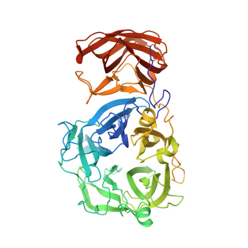
Structural analysis of a glycoside hydrolase family 43 arabinoxylan arabinofuranohydrolase in complex with xylotetraose reveals a different binding mechanism compared with other members of the same family.

Vandermarliere, E.Bourgois, T.M.Winn, M.D.van Campenhout, S.Volckaert, G.Delcour, J.A.Strelkov, S.V.Rabijns, A.Courtin, C.M.

(2009) Biochem J 418: 39-47

  • DOI: https://doi.org/10.1042/BJ20081256
  • Primary Citation of Related Structures:  
    3C7E, 3C7F, 3C7G, 3C7H, 3C7O

  • PubMed Abstract: 

    AXHs (arabinoxylan arabinofuranohydrolases) are alpha-L-arabinofuranosidases that specifically hydrolyse the glycosidic bond between arabinofuranosyl substituents and xylopyranosyl backbone residues of arabinoxylan. Bacillus subtilis was recently shown to produce an AXH that cleaves arabinose units from O-2- or O-3-mono-substituted xylose residues: BsAXH-m2,3 (B. subtilis AXH-m2,3). Crystallographic analysis reveals a two-domain structure for this enzyme: a catalytic domain displaying a five-bladed beta-propeller fold characteristic of GH (glycoside hydrolase) family 43 and a CBM (carbohydrate-binding module) with a beta-sandwich fold belonging to CBM family 6. Binding of substrate to BsAXH-m2,3 is largely based on hydrophobic stacking interactions, which probably allow the positional flexibility needed to hydrolyse both arabinose substituents at the O-2 or O-3 position of the xylose unit. Superposition of the BsAXH-m2,3 structure with known structures of the GH family 43 exo-acting enzymes, beta-xylosidase and alpha-L-arabinanase, each in complex with their substrate, reveals a different orientation of the sugar backbone.


  • Organizational Affiliation

    Laboratory for Biocrystallography, Department of Pharmaceutical Sciences, Katholieke Universiteit Leuven, Herestraat 49, O&N II, bus 822, 3000 Leuven, Belgium.


Macromolecules
Find similar proteins by:  (by identity cutoff)  |  3D Structure
Entity ID: 1
MoleculeChains Sequence LengthOrganismDetailsImage
Endo-1,4-beta-xylanase488Bacillus subtilisMutation(s): 0 
Gene Names: xynD
EC: 3.2.1.55
UniProt
Find proteins for Q45071 (Bacillus subtilis (strain 168))
Explore Q45071 
Go to UniProtKB:  Q45071
Entity Groups  
Sequence Clusters30% Identity50% Identity70% Identity90% Identity95% Identity100% Identity
UniProt GroupQ45071
Sequence Annotations
Expand
  • Reference Sequence
Oligosaccharides

Help

Entity ID: 2
MoleculeChains Length2D Diagram Glycosylation3D Interactions
beta-D-xylopyranose-(1-4)-beta-D-xylopyranose-(1-4)-beta-D-xylopyranose-(1-4)-beta-D-xylopyranose
B
4N/A
Glycosylation Resources
GlyTouCan:  G60650FG
GlyCosmos:  G60650FG
Experimental Data & Validation

Experimental Data

  • Method: X-RAY DIFFRACTION
  • Resolution: 2.02 Å
  • R-Value Free: 0.187 
  • R-Value Work: 0.143 
  • R-Value Observed: 0.145 
  • Space Group: P 21 21 21
Unit Cell:
Length ( Å )Angle ( ˚ )
a = 67.523α = 90
b = 72.401β = 90
c = 106.869γ = 90
Software Package:
Software NamePurpose
REFMACrefinement
DENZOdata reduction
SCALEPACKdata scaling
PHASERphasing

Structure Validation

View Full Validation Report


Entry History 

Revision History  (Full details and data files)

  • Version 1.0: 2008-11-18
    Type: Initial release
  • Version 1.1: 2011-07-13
    Changes: Non-polymer description, Version format compliance
  • Version 2.0: 2020-07-29
    Type: Remediation
    Reason: Carbohydrate remediation
    Changes: Advisory, Atomic model, Data collection, Database references, Derived calculations, Structure summary
  • Version 2.1: 2023-08-30
    Changes: Data collection, Database references, Derived calculations, Refinement description, Structure summary