3BX4

Crystal structure of the snake venom toxin aggretin


Experimental Data Snapshot

  • Method: X-RAY DIFFRACTION
  • Resolution: 1.70 Å
  • R-Value Free: 0.236 
  • R-Value Work: 0.202 
  • R-Value Observed: 0.204 

wwPDB Validation   3D Report Full Report


This is version 1.2 of the entry. See complete history


Literature

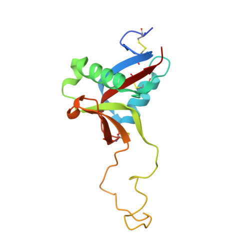
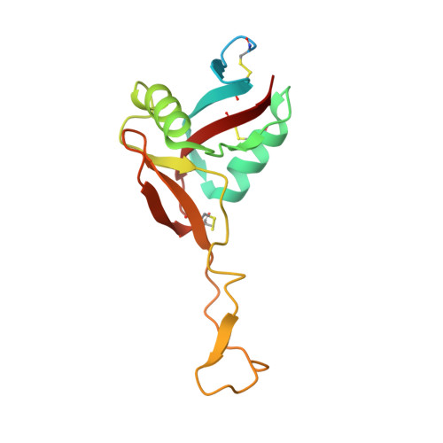
The crystal structure of the platelet activator aggretin reveals a novel (alphabeta)2 dimeric structure.

Hooley, E.Papagrigoriou, E.Navdaev, A.Pandey, A.V.Clemetson, J.M.Clemetson, K.J.Emsley, J.

(2008) Biochemistry 47: 7831-7837

  • DOI: https://doi.org/10.1021/bi800528t
  • Primary Citation of Related Structures:  
    3BX4

  • PubMed Abstract: 

    Aggretin is a C-type lectin purified from Calloselasma rhodostoma snake venom. It is a potent activator of platelets, resulting in a collagen-like response by binding and clustering platelet receptor CLEC-2. We present here the crystal structure of aggretin at 1.7 A which reveals a unique tetrameric quaternary structure. The two alphabeta heterodimers are arranged through 2-fold rotational symmetry, resulting in an antiparallel side-by-side arrangement. Aggretin thus presents two ligand binding sites on one surface and can therefore cluster ligands in a manner reminiscent of convulxin and flavocetin. To examine the molecular basis of the interaction with CLEC-2, we used a molecular modeling approach of docking the aggretin alphabeta structure with the CLEC-2 N-terminal domain (CLEC-2N). This model positions the CLEC-2N structure face down in the "saddle"-shaped binding site which lies between the aggretin alpha and beta lectin-like domains. A 2-fold rotation of this complex to generate the aggretin tetramer reveals dimer contacts for CLEC-2N which bring the N- and C-termini into the proximity of each other, and a series of contacts involving two interlocking beta-strands close to the N-terminus are described. A comparison with homologous lectin-like domains from the immunoreceptor family reveals a similar but not identical dimerization mode, suggesting this structure may represent the clustered form of CLEC-2 capable of signaling across the platelet membrane.


  • Organizational Affiliation

    Centre for Biomolecular Sciences, School of Pharmacy, University of Nottingham, UK.


Macromolecules
Find similar proteins by:  (by identity cutoff)  |  3D Structure
Entity ID: 1
MoleculeChains Sequence LengthOrganismDetailsImage
Aggretin alpha chain
A, C
136N/AMutation(s): 0 
UniProt
Find proteins for Q9I841 (Calloselasma rhodostoma)
Explore Q9I841 
Go to UniProtKB:  Q9I841
Entity Groups  
Sequence Clusters30% Identity50% Identity70% Identity90% Identity95% Identity100% Identity
UniProt GroupQ9I841
Sequence Annotations
Expand
  • Reference Sequence
Find similar proteins by:  (by identity cutoff)  |  3D Structure
Entity ID: 2
MoleculeChains Sequence LengthOrganismDetailsImage
Aggretin beta chain
B, D
146N/AMutation(s): 0 
UniProt
Find proteins for Q9I840 (Calloselasma rhodostoma)
Explore Q9I840 
Go to UniProtKB:  Q9I840
Entity Groups  
Sequence Clusters30% Identity50% Identity70% Identity90% Identity95% Identity100% Identity
UniProt GroupQ9I840
Sequence Annotations
Expand
  • Reference Sequence
Small Molecules
Ligands 2 Unique
IDChains Name / Formula / InChI Key2D Diagram3D Interactions
SO4
Query on SO4

Download Ideal Coordinates CCD File 
E [auth A]
F [auth A]
G [auth B]
H [auth B]
I [auth C]
E [auth A],
F [auth A],
G [auth B],
H [auth B],
I [auth C],
J [auth C],
K [auth D],
L [auth D],
M [auth D]
SULFATE ION
O4 S
QAOWNCQODCNURD-UHFFFAOYSA-L
GOL
Query on GOL

Download Ideal Coordinates CCD File 
N [auth D]GLYCEROL
C3 H8 O3
PEDCQBHIVMGVHV-UHFFFAOYSA-N
Experimental Data & Validation

Experimental Data

  • Method: X-RAY DIFFRACTION
  • Resolution: 1.70 Å
  • R-Value Free: 0.236 
  • R-Value Work: 0.202 
  • R-Value Observed: 0.204 
  • Space Group: P 21 21 2
Unit Cell:
Length ( Å )Angle ( ˚ )
a = 64.318α = 90
b = 91.305β = 90
c = 118.916γ = 90
Software Package:
Software NamePurpose
REFMACrefinement
DENZOdata reduction
SCALEPACKdata scaling
AMoREphasing

Structure Validation

View Full Validation Report


Entry History 

Deposition Data

Revision History  (Full details and data files)

  • Version 1.0: 2008-08-26
    Type: Initial release
  • Version 1.1: 2011-07-13
    Changes: Non-polymer description, Version format compliance
  • Version 1.2: 2023-08-30
    Changes: Data collection, Database references, Derived calculations, Refinement description