3BU7

Crystal Structure and Biochemical Characterization of GDOsp, a Gentisate 1,2-Dioxygenase from Silicibacter Pomeroyi


Experimental Data Snapshot

  • Method: X-RAY DIFFRACTION
  • Resolution: 2.80 Å
  • R-Value Free: 0.245 
  • R-Value Work: 0.210 
  • R-Value Observed: 0.212 

wwPDB Validation   3D Report Full Report


This is version 1.3 of the entry. See complete history


Literature

Crystal structure and mutagenic analysis of GDOsp, a gentisate 1,2-dioxygenase from Silicibacter pomeroyi.

Chen, J.Li, W.Wang, M.Zhu, G.Liu, D.Sun, F.Hao, N.Li, X.Rao, Z.Zhang, X.C.

(2008) Protein Sci 17: 1362-1373

  • DOI: https://doi.org/10.1110/ps.035881.108
  • Primary Citation of Related Structures:  
    3BU7

  • PubMed Abstract: 

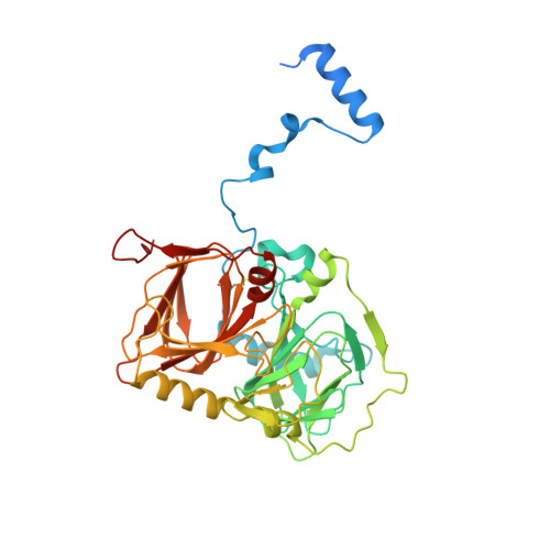
    Dioxygenases catalyze dioxygen incorporation into various organic compounds and play a key role in the complex degradation pathway of mono- and polycyclic aromatic and hetero-aromatic compounds. Here we report the crystal structure of gentisate 1,2-dioxygenase from Silicibacter pomeroyi (GDOsp) at a 2.8 A resolution. The enzyme possessed a conserved three-dimensional structure of the bicupin family, forming a homotetramerization. However, each subunit of GDOsp unusually contained two ferrous centers that were located in its two homologous cupin domains, respectively. Further mutagenic analysis indicated that the enzyme activity of GDOsp depends on the microenvironment in both metal-binding sites. Moreover, homologous structural comparison and functional study on GDOsp variants unveiled a group of functionally essential residues and suggested that the active site of the enzyme is located in the amino-terminal domain, but could be influenced by changes in the carboxyl domain. Therefore, GDOsp may provide a working model for studying long-distance communication within a protein (or its complex).


  • Organizational Affiliation

    National Laboratory of Biomacromolecules, Institute of Biophysics, Chinese Academy of Sciences, Beijing 100101, People's Republic of China.


Macromolecules
Find similar proteins by:  (by identity cutoff)  |  3D Structure
Entity ID: 1
MoleculeChains Sequence LengthOrganismDetailsImage
Gentisate 1,2-dioxygenase
A, B
394N/AMutation(s): 0 
Gene Names: gtdA-2
EC: 1.13.11.4
UniProt
Find proteins for Q5LLB1 (Ruegeria pomeroyi (strain ATCC 700808 / DSM 15171 / DSS-3))
Explore Q5LLB1 
Go to UniProtKB:  Q5LLB1
Entity Groups  
Sequence Clusters30% Identity50% Identity70% Identity90% Identity95% Identity100% Identity
UniProt GroupQ5LLB1
Sequence Annotations
Expand
  • Reference Sequence
Experimental Data & Validation

Experimental Data

  • Method: X-RAY DIFFRACTION
  • Resolution: 2.80 Å
  • R-Value Free: 0.245 
  • R-Value Work: 0.210 
  • R-Value Observed: 0.212 
  • Space Group: H 3 2
Unit Cell:
Length ( Å )Angle ( ˚ )
a = 129.999α = 90
b = 129.999β = 90
c = 246.255γ = 120
Software Package:
Software NamePurpose
DENZOdata reduction
SCALEPACKdata scaling
MOLREPphasing
REFMACrefinement
PDB_EXTRACTdata extraction
CrystalCleardata collection
HKL-2000data reduction

Structure Validation

View Full Validation Report


Entry History 

Deposition Data

Revision History  (Full details and data files)

  • Version 1.0: 2008-08-12
    Type: Initial release
  • Version 1.1: 2011-07-13
    Changes: Version format compliance
  • Version 1.2: 2017-10-25
    Changes: Refinement description
  • Version 1.3: 2023-08-30
    Changes: Data collection, Database references, Derived calculations, Refinement description