3BBT

crystal structure of the ErbB4 kinase in complex with lapatinib


Experimental Data Snapshot

  • Method: X-RAY DIFFRACTION
  • Resolution: 2.80 Å
  • R-Value Free: 0.289 
  • R-Value Work: 0.251 
  • R-Value Observed: 0.253 

wwPDB Validation   3D Report Full Report


This is version 1.3 of the entry. See complete history


Literature

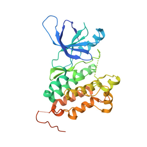
Mechanism of Activation and Inhibition of the HER4/ErbB4 Kinase.

Qiu, C.Tarrant, M.K.Choi, S.H.Sathyamurthy, A.Bose, R.Banjade, S.Pal, A.Bornmann, W.G.Lemmon, M.A.Cole, P.A.Leahy, D.J.

(2008) Structure 16: 460-467

  • DOI: https://doi.org/10.1016/j.str.2007.12.016
  • Primary Citation of Related Structures:  
    3BBT, 3BBW, 3BCE

  • PubMed Abstract: 

    HER4/ErbB4 is a ubiquitously expressed member of the EGF/ErbB family of receptor tyrosine kinases that is essential for normal development of the heart, nervous system, and mammary gland. We report here crystal structures of the ErbB4 kinase domain in active and lapatinib-inhibited forms. Active ErbB4 kinase adopts an asymmetric dimer conformation essentially identical to that observed to be important for activation of the EGF receptor/ErbB1 kinase. Mutagenesis studies of intact ErbB4 in Ba/F3 cells confirm the importance of this asymmetric dimer for activation of intact ErbB4. Lapatinib binds to an inactive form of the ErbB4 kinase in a mode equivalent to its interaction with the EGF receptor. All ErbB4 residues contacted by lapatinib are conserved in the EGF receptor and HER2/ErbB2, which lapatinib also targets. These results demonstrate that key elements of kinase activation and inhibition are conserved among ErbB family members.


  • Organizational Affiliation

    Department of Biophysics & Biophysical Chemistry, Johns Hopkins University School of Medicine, Baltimore, MD 21205, USA.


Macromolecules
Find similar proteins by:  (by identity cutoff)  |  3D Structure
Entity ID: 1
MoleculeChains Sequence LengthOrganismDetailsImage
Receptor tyrosine-protein kinase erbB-4A [auth B],
B [auth D]
328Homo sapiensMutation(s): 0 
Gene Names: ERBB4HER4
EC: 2.7.10.1
UniProt & NIH Common Fund Data Resources
Find proteins for Q15303 (Homo sapiens)
Explore Q15303 
Go to UniProtKB:  Q15303
PHAROS:  Q15303
GTEx:  ENSG00000178568 
Entity Groups  
Sequence Clusters30% Identity50% Identity70% Identity90% Identity95% Identity100% Identity
UniProt GroupQ15303
Sequence Annotations
Expand
  • Reference Sequence
Small Molecules
Ligands 1 Unique
IDChains Name / Formula / InChI Key2D Diagram3D Interactions
FMM
Query on FMM

Download Ideal Coordinates CCD File 
C [auth B],
D
N-{3-CHLORO-4-[(3-FLUOROBENZYL)OXY]PHENYL}-6-[5-({[2-(METHYLSULFONYL)ETHYL]AMINO}METHYL)-2-FURYL]-4-QUINAZOLINAMINE
C29 H26 Cl F N4 O4 S
BCFGMOOMADDAQU-UHFFFAOYSA-N
Binding Affinity Annotations 
IDSourceBinding Affinity
FMM PDBBind:  3BBT Ki: 700 (nM) from 1 assay(s)
BindingDB:  3BBT Kd: 54 (nM) from 1 assay(s)
IC50: min: 7.6, max: 1000 (nM) from 6 assay(s)
Experimental Data & Validation

Experimental Data

  • Method: X-RAY DIFFRACTION
  • Resolution: 2.80 Å
  • R-Value Free: 0.289 
  • R-Value Work: 0.251 
  • R-Value Observed: 0.253 
  • Space Group: P 61
Unit Cell:
Length ( Å )Angle ( ˚ )
a = 102.682α = 90
b = 102.682β = 90
c = 185.124γ = 120
Software Package:
Software NamePurpose
DENZOdata reduction
SCALEPACKdata scaling
PHASERphasing
REFMACrefinement
PDB_EXTRACTdata extraction

Structure Validation

View Full Validation Report


Entry History 

Deposition Data

  • Released Date: 2008-02-12 
  • Deposition Author(s): Qiu, C.

Revision History  (Full details and data files)

  • Version 1.0: 2008-02-12
    Type: Initial release
  • Version 1.1: 2011-07-13
    Changes: Advisory, Refinement description, Version format compliance
  • Version 1.2: 2017-10-25
    Changes: Refinement description
  • Version 1.3: 2024-02-21
    Changes: Data collection, Database references, Derived calculations, Refinement description