


The crystal structure of mannonate dehydratase from Streptococcus suis serotype2
Peng, H., Zhang, Q.M., Gao, F., Liu, Y.W., Qi, J.X., Gao, G.F.To be published.
Experimental Data Snapshot
wwPDB Validation 3D Report Full Report

Entity ID: 1 | |||||
|---|---|---|---|---|---|
| Molecule | Chains | Sequence Length | Organism | Details | Image |
| D-mannonate dehydratase | 386 | Streptococcus suis | Mutation(s): 0 Gene Names: UxuA EC: 4.2.1.8 | 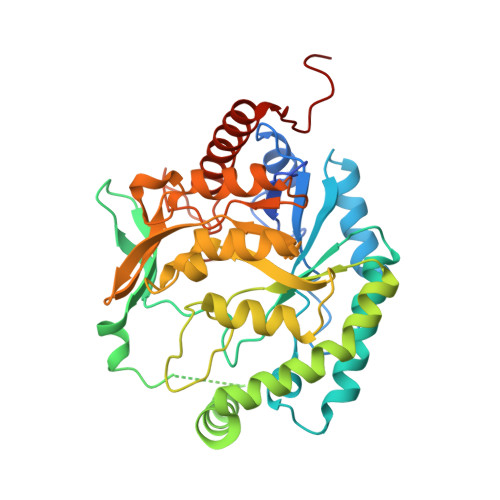 | |
UniProt | |||||
Find proteins for A4VVI4 (Streptococcus suis (strain 05ZYH33)) Explore A4VVI4 Go to UniProtKB: A4VVI4 | |||||
Entity Groups | |||||
| Sequence Clusters | 30% Identity50% Identity70% Identity90% Identity95% Identity100% Identity | ||||
| UniProt Group | A4VVI4 | ||||
Sequence AnnotationsExpand | |||||
| |||||
| Length ( Å ) | Angle ( ˚ ) |
|---|---|
| a = 105.69 | α = 90 |
| b = 105.69 | β = 90 |
| c = 159.572 | γ = 90 |
| Software Name | Purpose |
|---|---|
| REFMAC | refinement |
| CrystalClear | data collection |
| CrystalClear | data reduction |
| CrystalClear | data scaling |
| MOLREP | phasing |
RCSB PDB (citation) is hosted by
RCSB PDB is a member of the

