Structural and mutational analyses of the receptor binding domain of botulinum D/C mosaic neurotoxin: insight into the ganglioside binding mechanism
Nuemket, N., Tanaka, Y., Tsukamoto, K., Tsuji, T., Nakamura, K., Kozaki, S., Yao, M., Tanaka, I.(2011) Biochem Biophys Res Commun 411: 433-439
- PubMed: 21749855
- DOI: https://doi.org/10.1016/j.bbrc.2011.06.173
- Primary Citation of Related Structures:
3AZV, 3AZW - PubMed Abstract:
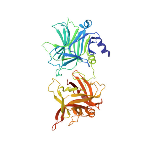
Clostridium botulinum type D strain OFD05, which produces the D/C mosaic neurotoxin, was isolated from cattle killed by the recent botulism outbreak in Japan. The D/C mosaic neurotoxin is the most toxic of the botulinum neurotoxins (BoNT) characterized to date. Here, we determined the crystal structure of the receptor binding domain of BoNT from strain OFD05 in complex with 3'-sialyllactose at a resolution of 3.0Å. In the structure, an electron density derived from the 3'-sialyllactose was confirmed at the cleft in the C-terminal subdomain. Alanine site-directed mutagenesis showed the significant contribution of the residues surrounding the cleft to ganglioside recognition. In addition, a loop adjoining the cleft also plays an important role in ganglioside recognition. In contrast, little effect was observed when the residues located around the surface previously identified as the protein receptor binding site in other BoNTs were substituted. The results of cell binding analysis of the mutants were significantly correlated with the ganglioside binding properties. Based on these observations, a cell binding mechanism of BoNT from strain OFD05 is proposed, which involves cooperative contribution of two ganglioside binding sites.
Organizational Affiliation:
Graduate School of Life Sciences, Hokkaido University, Sapporo 060-0810, Japan.