3ASJ

Crystal structure of homoisocitrate dehydrogenase in complex with a designed inhibitor


Experimental Data Snapshot

  • Method: X-RAY DIFFRACTION
  • Resolution: 2.60 Å
  • R-Value Free: 0.231 
  • R-Value Work: 0.169 
  • R-Value Observed: 0.172 

wwPDB Validation   3D Report Full Report


Ligand Structure Quality Assessment 


This is version 1.2 of the entry. See complete history


Literature

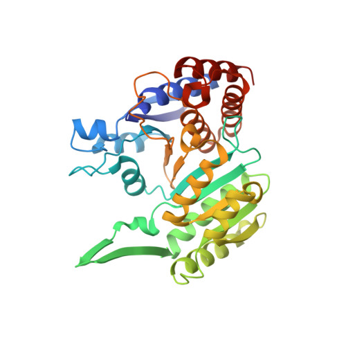
Structure of Thermus thermophilus homoisocitrate dehydrogenase in complex with a designed inhibitor

Nango, E.Yamamoto, T.Kumasaka, T.Eguchi, T.

(2011) J Biochem 150: 607-614

  • DOI: https://doi.org/10.1093/jb/mvr097
  • Primary Citation of Related Structures:  
    3ASJ

  • PubMed Abstract: 

    Homoisocitrate dehydrogenase (HICDH) is involved in the α-aminoadipate pathway of lysine biosynthesis in some bacteria and higher fungi, and catalyses the oxidative decarboxylation of (2R,3S)-homoisocitrate into 2-ketoadipate using NAD(+) as a coenzyme. In this study, the crystal structure of Thermus thermophilus HICDH in a binary complex with a designed inhibitor, (2S,3S)-thiahomoisocitrate, has been determined at 2.6 Å resolution. The inhibitor observed as a decarboxylated product interacts through hydrogen bonding to Arg 118, Tyr 125 and Lys 171 in the active site. The induced fitting was also observed around the region consisting of residues 120-141, which shifted up to 2.8 Å towards the active site. In addition, it was found that the complex structure adopts a closed conformation in two domains. While the structure of apo-HICDH shows that a catalytic residue Tyr 125 and Arg 85 that engages in substrate recognition are flipped out of the active site, these residues turn towards the active site in the complex structure. The results revealed that they directly interact with a substrate and are involved in catalysis or substrate recognition. Furthermore, by comparing the binary complex with the quaternary complex of Escherichia coli isocitrate dehydrogenase, the substrate recognition mechanism of HICDH is also discussed.


  • Organizational Affiliation

    Department of Chemistry, Tokyo Institute of Technology, Tokyo Institute of Technology, O-okayama, Meguro-ku, Tokyo 152-8551, Japan.


Macromolecules
Find similar proteins by:  (by identity cutoff)  |  3D Structure
Entity ID: 1
MoleculeChains Sequence LengthOrganismDetailsImage
Homoisocitrate dehydrogenase
A, B, C, D
334Thermus thermophilusMutation(s): 0 
Gene Names: hicdhdh
UniProt
Find proteins for Q5SIJ1 (Thermus thermophilus (strain ATCC 27634 / DSM 579 / HB8))
Explore Q5SIJ1 
Go to UniProtKB:  Q5SIJ1
Entity Groups  
Sequence Clusters30% Identity50% Identity70% Identity90% Identity95% Identity100% Identity
UniProt GroupQ5SIJ1
Sequence Annotations
Expand
  • Reference Sequence
Small Molecules
Ligands 3 Unique
IDChains Name / Formula / InChI Key2D Diagram3D Interactions
XYM
Query on XYM

Download Ideal Coordinates CCD File 
K [auth B],
W [auth D]
(2Z)-3-[(carboxymethyl)sulfanyl]-2-hydroxyprop-2-enoic acid
C5 H6 O5 S
IXLZHTPCCBDKBW-IWQZZHSRSA-N
XYN
Query on XYN

Download Ideal Coordinates CCD File 
E [auth A]3-[(carboxymethyl)sulfanyl]-2-oxopropanoic acid
C5 H6 O5 S
FDUKYMHTXNDQPD-UHFFFAOYSA-N
MPD
Query on MPD

Download Ideal Coordinates CCD File 
F [auth A]
G [auth A]
H [auth A]
I [auth A]
J [auth A]
F [auth A],
G [auth A],
H [auth A],
I [auth A],
J [auth A],
L [auth B],
M [auth B],
N [auth B],
O [auth B],
P [auth B],
Q [auth B],
R [auth B],
S [auth C],
T [auth C],
U [auth C],
V [auth C],
X [auth D],
Y [auth D],
Z [auth D]
(4S)-2-METHYL-2,4-PENTANEDIOL
C6 H14 O2
SVTBMSDMJJWYQN-YFKPBYRVSA-N
Experimental Data & Validation

Experimental Data

  • Method: X-RAY DIFFRACTION
  • Resolution: 2.60 Å
  • R-Value Free: 0.231 
  • R-Value Work: 0.169 
  • R-Value Observed: 0.172 
  • Space Group: C 1 2 1
Unit Cell:
Length ( Å )Angle ( ˚ )
a = 220.967α = 90
b = 93.695β = 97.68
c = 88.802γ = 90
Software Package:
Software NamePurpose
BSSdata collection
MOLREPphasing
REFMACrefinement
HKL-2000data reduction
HKL-2000data scaling

Structure Validation

View Full Validation Report


Ligand Structure Quality Assessment 


Entry History 

Deposition Data

Revision History  (Full details and data files)

  • Version 1.0: 2011-09-21
    Type: Initial release
  • Version 1.1: 2013-06-19
    Changes: Database references
  • Version 1.2: 2023-11-01
    Changes: Data collection, Database references, Derived calculations, Refinement description