3AJZ

Crystal Structure of Ancestral Congerin Con-anc


Experimental Data Snapshot

  • Method: X-RAY DIFFRACTION
  • Resolution: 1.50 Å
  • R-Value Free: 0.217 
  • R-Value Work: 0.204 
  • R-Value Observed: 0.204 

wwPDB Validation   3D Report Full Report


This is version 2.1 of the entry. See complete history


Literature

Tracing protein evolution through ancestral structures of fish galectin

Konno, A.Kitagawa, A.Watanabe, M.Ogawa, T.Shirai, T.

(2011) Structure 19: 711-721

  • DOI: https://doi.org/10.1016/j.str.2011.02.014
  • Primary Citation of Related Structures:  
    3AJY, 3AJZ, 3AK0

  • PubMed Abstract: 

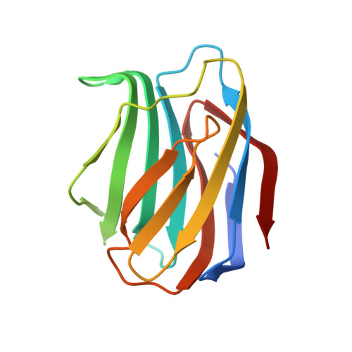
    Ancestral structures of fish galectins (congerins) were determined. The extant isoforms I and II of congerin are the components of a fish biological defense system and have rapidly differentiated under natural selection pressure, by which congerin I has experienced a protein-fold evolution. The dimer structure of the ancestral congerin demonstrated intermediate features of the extant isoforms. The protein-fold evolution was not observed in the ancestral structure, indicating it specifically occurred in congerin I lineage. Details of hydrogen bonding pattern at the dimer interface and the carbohydrate-binding site of the ancestor were different from the current proteins. The differences implied these proteins were under selection pressure for stabilizing dimer structure and differentiation in carbohydrate specificity. The ancestor had rather low cytotoxic activity than offspring, indicating selection was made to enhance this activity of congerins. Combined with functional analyses, the structure revealed atomic details of the differentiation process of the proteins.


  • Organizational Affiliation

    Department of Biomolecular Science, Graduate School of Life Sciences, Tohoku University, Sendai 980-8577, Japan.


Macromolecules
Find similar proteins by:  (by identity cutoff)  |  3D Structure
Entity ID: 1
MoleculeChains Sequence LengthOrganismDetailsImage
Ancestral congerin Con-anc135N/AMutation(s): 0 
Entity Groups  
Sequence Clusters30% Identity50% Identity70% Identity90% Identity95% Identity100% Identity
Sequence Annotations
Expand
  • Reference Sequence
Oligosaccharides

Help

Entity ID: 2
MoleculeChains Length2D Diagram Glycosylation3D Interactions
beta-D-galactopyranose-(1-4)-beta-D-glucopyranose
B
2N/A
Glycosylation Resources
GlyTouCan:  G84224TW
GlyCosmos:  G84224TW
GlyGen:  G84224TW
Biologically Interesting Molecules (External Reference) 1 Unique
Experimental Data & Validation

Experimental Data

  • Method: X-RAY DIFFRACTION
  • Resolution: 1.50 Å
  • R-Value Free: 0.217 
  • R-Value Work: 0.204 
  • R-Value Observed: 0.204 
  • Space Group: I 2 2 2
Unit Cell:
Length ( Å )Angle ( ˚ )
a = 53.908α = 90
b = 77.519β = 90
c = 88.374γ = 90
Software Package:
Software NamePurpose
HKL-2000data collection
MOLREPphasing
REFMACrefinement
HKL-2000data reduction
SCALEPACKdata scaling

Structure Validation

View Full Validation Report


Entry History 

Deposition Data

Revision History  (Full details and data files)

  • Version 1.0: 2011-05-18
    Type: Initial release
  • Version 1.1: 2011-07-13
    Changes: Version format compliance
  • Version 1.2: 2013-08-07
    Changes: Database references
  • Version 1.3: 2015-02-11
    Changes: Derived calculations
  • Version 2.0: 2020-07-29
    Type: Remediation
    Reason: Carbohydrate remediation
    Changes: Atomic model, Data collection, Database references, Derived calculations, Structure summary
  • Version 2.1: 2023-11-01
    Changes: Data collection, Database references, Refinement description, Structure summary