3AJH

Crystal structure of PcyA V225D-biliverdin XIII alpha complex


Experimental Data Snapshot

  • Method: X-RAY DIFFRACTION
  • Resolution: 2.25 Å
  • R-Value Free: 0.289 
  • R-Value Work: 0.251 
  • R-Value Observed: 0.251 

wwPDB Validation   3D Report Full Report


Ligand Structure Quality Assessment 


This is version 1.2 of the entry. See complete history


Literature

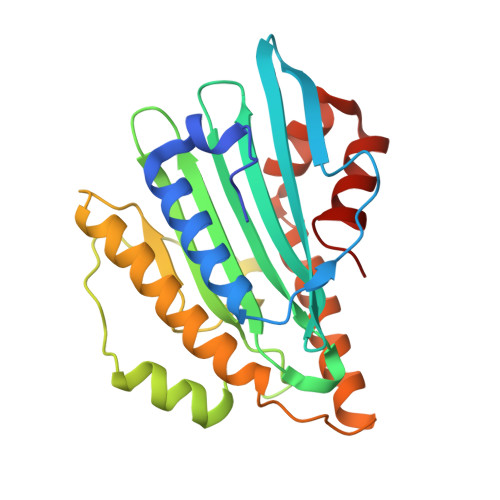
One residue substitution in PcyA leads to unexpected changes in tetrapyrrole substrate binding.

Wada, K.Hagiwara, Y.Yutani, Y.Fukuyama, K.

(2010) Biochem Biophys Res Commun 402: 373-377

  • DOI: https://doi.org/10.1016/j.bbrc.2010.10.037
  • Primary Citation of Related Structures:  
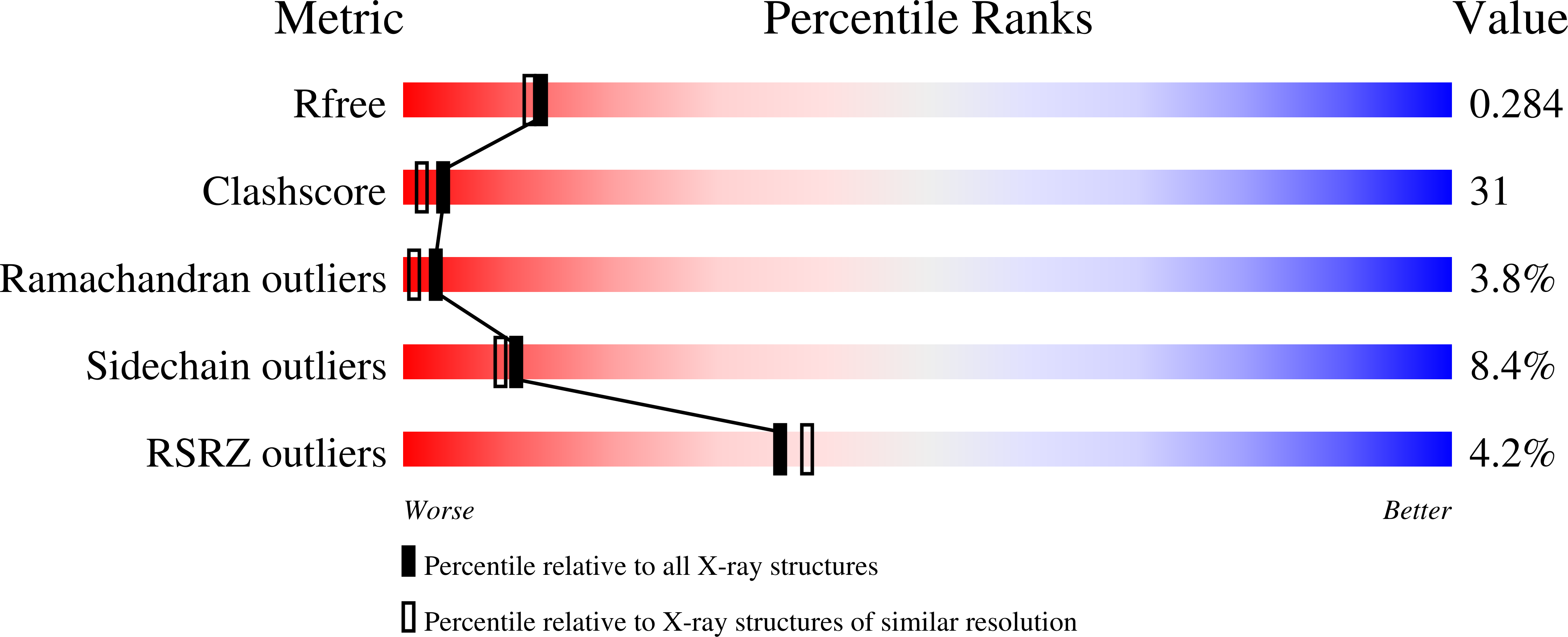
    3AJG, 3AJH

  • PubMed Abstract: 

    Phycocyanobilin:ferredoxin oxidoreductase (PcyA) catalyzes the sequential reduction of the vinyl group of the D-ring and A-ring of biliverdin IXα (BV), using reducing equivalents provided by ferredoxin. This reaction produces phycocyanobilin, a pigment used for light-harvesting and light-sensing in red algae and cyanobacteria. The crystal structure of PcyA-BV reveals that BV is specifically bound in the PcyA active pocket through extensive hydrophobic and hydrophilic interactions. During the course of a mutational study of PcyA, we observed that mutation of the V225 position, apart from the processing sites, conferred an unusual property on PcyA; V225D mutant protein could bind BV and its analog BV13, but these complexes showed a distinct UV-vis absorption spectrum from that of the wild-type PcyA-BV complex. The crystal structures of BV- and BV13-bound forms of V225D protein revealed that gross structural changes occurred near the substrate-binding pocket, and that the BV/BV13 binding manner in the pocket was dramatically altered. Protein folding in V225D-BV/BV13 was more similar to that of substrate-free PcyA than that in PcyA-BV; the "induced-fit" did not occur when BV/BV13 was bound to the V225D protein. The unexpected structural change presented here provides a cautionary note about interpreting functional data derived from a mutated protein in the absence of its exact structure.


  • Organizational Affiliation

    Department of Biological Sciences, Graduate School of Science, Osaka University, Toyonaka, Osaka 560-0043, Japan.


Macromolecules
Find similar proteins by:  (by identity cutoff)  |  3D Structure
Entity ID: 1
MoleculeChains Sequence LengthOrganismDetailsImage
Phycocyanobilin:ferredoxin oxidoreductase
A, B
248Synechocystis sp. PCC 6803Mutation(s): 1 
Gene Names: pcyAslr0116
EC: 1.3.7.5
UniProt
Find proteins for Q55891 (Synechocystis sp. (strain PCC 6803 / Kazusa))
Explore Q55891 
Go to UniProtKB:  Q55891
Entity Groups  
Sequence Clusters30% Identity50% Identity70% Identity90% Identity95% Identity100% Identity
UniProt GroupQ55891
Sequence Annotations
Expand
  • Reference Sequence
Small Molecules
Ligands 1 Unique
IDChains Name / Formula / InChI Key2D Diagram3D Interactions
BL3
Query on BL3

Download Ideal Coordinates CCD File 
C [auth A],
D [auth B]
3-[2-[(Z)-[3-(2-carboxyethyl)-5-[(Z)-(3-ethenyl-4-methyl-5-oxo-pyrrol-2-ylidene)methyl]-4-methyl-pyrrol-2-ylidene]methy l]-5-[(Z)-(3-ethenyl-4-methyl-5-oxo-pyrrol-2-ylidene)methyl]-4-methyl-1H-pyrrol-3-yl]propanoic acid
C33 H34 N4 O6
JXYQCWFZZAWTEC-SRVCBVSDSA-N
Experimental Data & Validation

Experimental Data

  • Method: X-RAY DIFFRACTION
  • Resolution: 2.25 Å
  • R-Value Free: 0.289 
  • R-Value Work: 0.251 
  • R-Value Observed: 0.251 
  • Space Group: P 21 21 21
Unit Cell:
Length ( Å )Angle ( ˚ )
a = 74.611α = 90
b = 74.605β = 90
c = 84.627γ = 90
Software Package:
Software NamePurpose
HKL-2000data collection
MOLREPphasing
CNSrefinement
HKL-2000data reduction
HKL-2000data scaling

Structure Validation

View Full Validation Report


Ligand Structure Quality Assessment 


Entry History 

Deposition Data

Revision History  (Full details and data files)

  • Version 1.0: 2011-03-16
    Type: Initial release
  • Version 1.1: 2011-07-13
    Changes: Version format compliance
  • Version 1.2: 2024-03-13
    Changes: Data collection, Database references, Derived calculations, Structure summary