3A79

Crystal structure of TLR2-TLR6-Pam2CSK4 complex


Experimental Data Snapshot

  • Method: X-RAY DIFFRACTION
  • Resolution: 2.90 Å
  • R-Value Free: 0.280 
  • R-Value Work: 0.208 
  • R-Value Observed: 0.215 

wwPDB Validation   3D Report Full Report


Ligand Structure Quality Assessment 


This is version 2.1 of the entry. See complete history


Literature

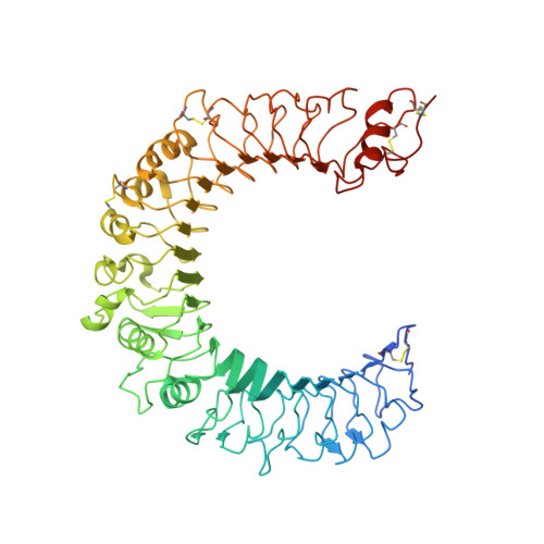
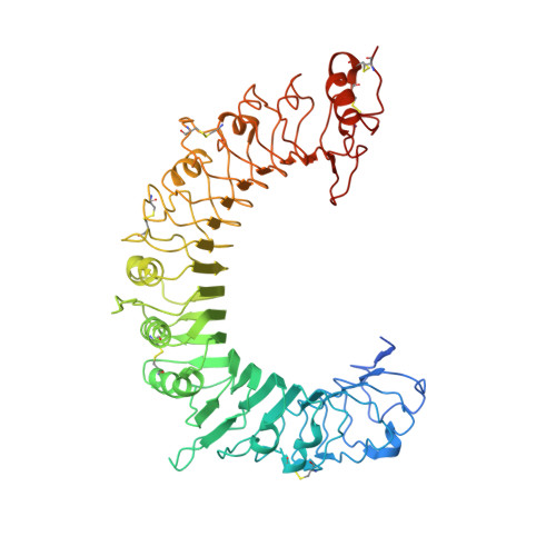
Recognition of lipopeptide patterns by Toll-like receptor 2-Toll-like receptor 6 heterodimer

Kang, J.Y.Nan, X.Jin, M.S.Youn, S.-J.Ryu, Y.H.Mah, S.Han, S.H.Lee, H.Paik, S.-G.Lee, J.-O.

(2009) Immunity 31: 873-884

  • DOI: https://doi.org/10.1016/j.immuni.2009.09.018
  • Primary Citation of Related Structures:  
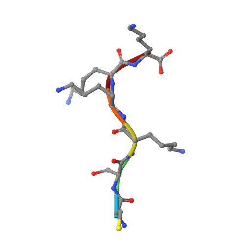
    3A79, 3A7B, 3A7C

  • PubMed Abstract: 

    Toll-like receptor 2 (TLR2) initiates potent immune responses by recognizing diacylated and triacylated lipopeptides. Its ligand specificity is controlled by whether it heterodimerizes with TLR1 or TLR6. We have determined the crystal structures of TLR2-TLR6-diacylated lipopeptide, TLR2-lipoteichoic acid, and TLR2-PE-DTPA complexes. PE-DTPA, 1,2-dimyristoyl-sn-glycero-3-phosphoethanolamine-N-diethylenetriaminepentaacetic acid, is a synthetic phospholipid derivative. Two major factors contribute to the ligand specificity of TLR2-TLR1 or TLR2-TLR6 heterodimers. First, the lipid channel of TLR6 is blocked by two phenylalanines. Simultaneous mutation of these phenylalanines made TLR2-TLR6 fully responsive not only to diacylated but also to triacylated lipopeptides. Second, the hydrophobic dimerization interface of TLR2-TLR6 is increased by 80%, which compensates for the lack of amide lipid interaction between the lipopeptide and TLR2-TLR6. The structures of the TLR2-lipoteichoic acid and the TLR2-PE-DTPA complexes demonstrate that a precise interaction pattern of the head group is essential for a robust immune response by TLR2 heterodimers.


  • Organizational Affiliation

    Department of Chemistry, KAIST, Daejon, 305-701, Korea.


Macromolecules
Find similar proteins by:  (by identity cutoff)  |  3D Structure
Entity ID: 1
MoleculeChains Sequence LengthOrganismDetailsImage
Toll-like receptor 2, Variable lymphocyte receptor B580Mus musculusEptatretus burgeri
This entity is chimeric
Mutation(s): 0 
Gene Names: Tlr2
UniProt
Find proteins for Q9QUN7 (Mus musculus)
Explore Q9QUN7 
Go to UniProtKB:  Q9QUN7
Find proteins for Q4G1L2 (Eptatretus burgeri)
Explore Q4G1L2 
Go to UniProtKB:  Q4G1L2
Entity Groups  
Sequence Clusters30% Identity50% Identity70% Identity90% Identity95% Identity100% Identity
UniProt GroupsQ4G1L2Q9QUN7
Sequence Annotations
Expand
  • Reference Sequence
Find similar proteins by:  (by identity cutoff)  |  3D Structure
Entity ID: 2
MoleculeChains Sequence LengthOrganismDetailsImage
Toll-like receptor 6, Variable lymphocyte receptor B562Mus musculusEptatretus burgeri
This entity is chimeric
Mutation(s): 0 
Gene Names: Tlr6
UniProt
Find proteins for Q4G1L3 (Eptatretus burgeri)
Explore Q4G1L3 
Go to UniProtKB:  Q4G1L3
Find proteins for Q9EPW9 (Mus musculus)
Explore Q9EPW9 
Go to UniProtKB:  Q9EPW9
Entity Groups  
Sequence Clusters30% Identity50% Identity70% Identity90% Identity95% Identity100% Identity
UniProt GroupsQ4G1L3Q9EPW9
Sequence Annotations
Expand
  • Reference Sequence

Find similar proteins by:  Sequence   |   3D Structure  

Entity ID: 3
MoleculeChains Sequence LengthOrganismDetailsImage
Pam2CSK46synthetic constructMutation(s): 0 
Sequence Annotations
Expand
  • Reference Sequence
Oligosaccharides

Help

Entity ID: 4
MoleculeChains Length2D Diagram Glycosylation3D Interactions
2-acetamido-2-deoxy-beta-D-glucopyranose-(1-4)-2-acetamido-2-deoxy-beta-D-glucopyranose
D, E, I
2N-Glycosylation
Glycosylation Resources
GlyTouCan:  G42666HT
GlyCosmos:  G42666HT
GlyGen:  G42666HT
Entity ID: 5
MoleculeChains Length2D Diagram Glycosylation3D Interactions
beta-D-mannopyranose-(1-4)-2-acetamido-2-deoxy-beta-D-glucopyranose-(1-4)-2-acetamido-2-deoxy-beta-D-glucopyranose
F, G, H
3N-Glycosylation
Glycosylation Resources
GlyTouCan:  G15407YE
GlyCosmos:  G15407YE
GlyGen:  G15407YE
Experimental Data & Validation

Experimental Data

  • Method: X-RAY DIFFRACTION
  • Resolution: 2.90 Å
  • R-Value Free: 0.280 
  • R-Value Work: 0.208 
  • R-Value Observed: 0.215 
  • Space Group: P 61 2 2
Unit Cell:
Length ( Å )Angle ( ˚ )
a = 168.901α = 90
b = 168.901β = 90
c = 231.353γ = 120
Software Package:
Software NamePurpose
PHENIXrefinement
PDB_EXTRACTdata extraction
XDSdata reduction
XSCALEdata scaling
PHASERphasing

Structure Validation

View Full Validation Report


Ligand Structure Quality Assessment 


Entry History 

Deposition Data

Revision History  (Full details and data files)

  • Version 1.0: 2009-11-24
    Type: Initial release
  • Version 1.1: 2011-07-13
    Changes: Non-polymer description, Version format compliance
  • Version 1.2: 2012-06-27
    Changes: Non-polymer description
  • Version 1.3: 2017-08-09
    Changes: Source and taxonomy
  • Version 1.4: 2017-08-16
    Changes: Source and taxonomy
  • Version 2.0: 2020-07-29
    Type: Remediation
    Reason: Carbohydrate remediation
    Changes: Advisory, Atomic model, Data collection, Database references, Derived calculations, Non-polymer description, Structure summary
  • Version 2.1: 2023-11-01
    Changes: Data collection, Database references, Refinement description, Structure summary