


Crystal structure of isomaltase from Saccharomyces cerevisiae
Yamamoto, K., Miyake, H., Kusunoki, M., Osaki, S.To be published.
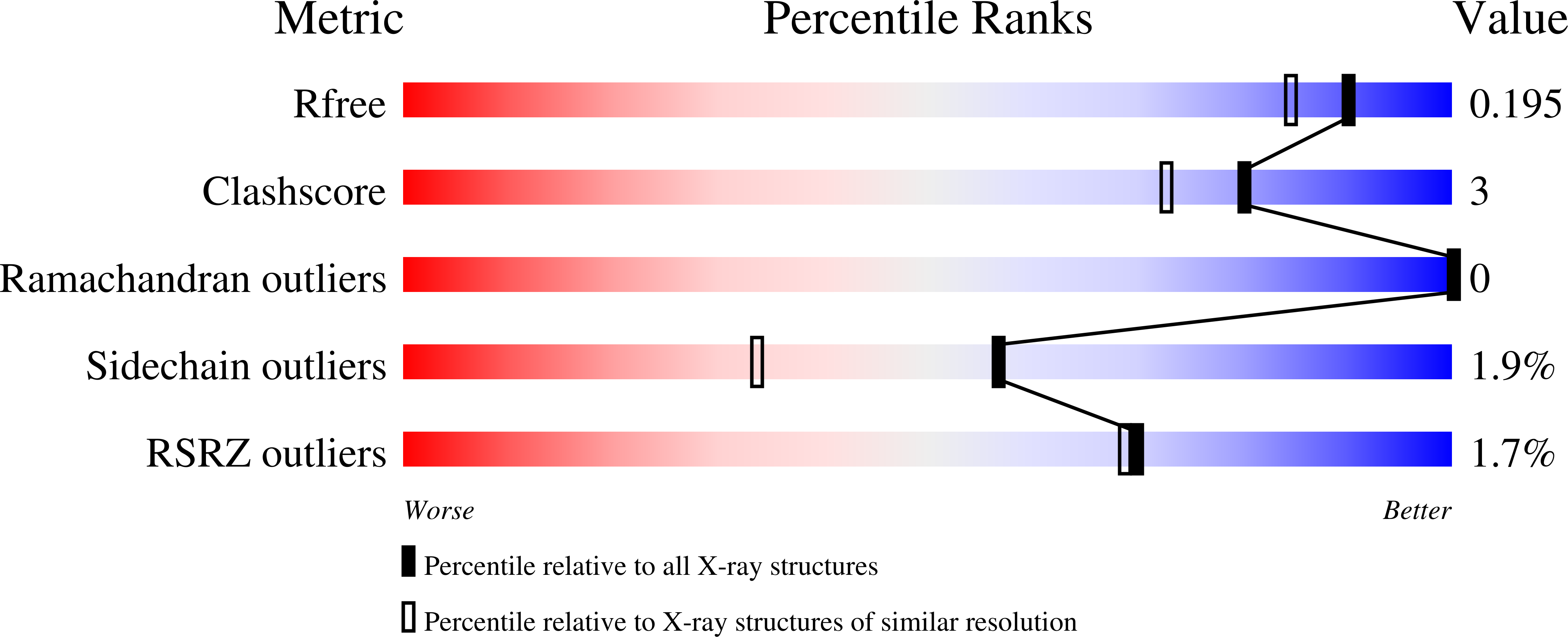
Experimental Data Snapshot
wwPDB Validation 3D Report Full Report
Entity ID: 1 | |||||
|---|---|---|---|---|---|
| Molecule | Chains | Sequence Length | Organism | Details | Image |
| Oligo-1,6-glucosidase | 589 | Saccharomyces cerevisiae | Mutation(s): 0 EC: 3.2.1.10 |  | |
UniProt | |||||
Find proteins for P53051 (Saccharomyces cerevisiae (strain ATCC 204508 / S288c)) Explore P53051 Go to UniProtKB: P53051 | |||||
Entity Groups | |||||
| Sequence Clusters | 30% Identity50% Identity70% Identity90% Identity95% Identity100% Identity | ||||
| UniProt Group | P53051 | ||||
Sequence AnnotationsExpand | |||||
| |||||
| Ligands 1 Unique | |||||
|---|---|---|---|---|---|
| ID | Chains | Name / Formula / InChI Key | 2D Diagram | 3D Interactions | |
| CA Query on CA | B [auth A] | CALCIUM ION Ca BHPQYMZQTOCNFJ-UHFFFAOYSA-N |  | ||
| Length ( Å ) | Angle ( ˚ ) |
|---|---|
| a = 95.709 | α = 90 |
| b = 115.674 | β = 91.33 |
| c = 61.837 | γ = 90 |
| Software Name | Purpose |
|---|---|
| REFMAC | refinement |
| HKL-2000 | data collection |
| HKL-2000 | data reduction |
| HKL-2000 | data scaling |
| MOLREP | phasing |
RCSB PDB (citation) is hosted by
RCSB PDB is a member of the

