3A3K

Reversibly bound chloride in the atrial natriuretic peptide receptor hormone-binding domain


Experimental Data Snapshot

  • Method: X-RAY DIFFRACTION
  • Resolution: 2.50 Å
  • R-Value Free: 0.243 
  • R-Value Work: 0.205 
  • R-Value Observed: 0.207 

wwPDB Validation   3D Report Full Report


Ligand Structure Quality Assessment 


This is version 2.0 of the entry. See complete history


Literature

Reversibly bound chloride in the atrial natriuretic peptide receptor hormone-binding domain: Possible allosteric regulation and a conserved structural motif for the chloride-binding site.

Ogawa, H.Qiu, Y.Philo, J.S.Arakawa, T.Ogata, C.M.Misono, K.S.

(2010) Protein Sci 19: 544-557

  • DOI: https://doi.org/10.1002/pro.332
  • Primary Citation of Related Structures:  
    3A3K

  • PubMed Abstract: 

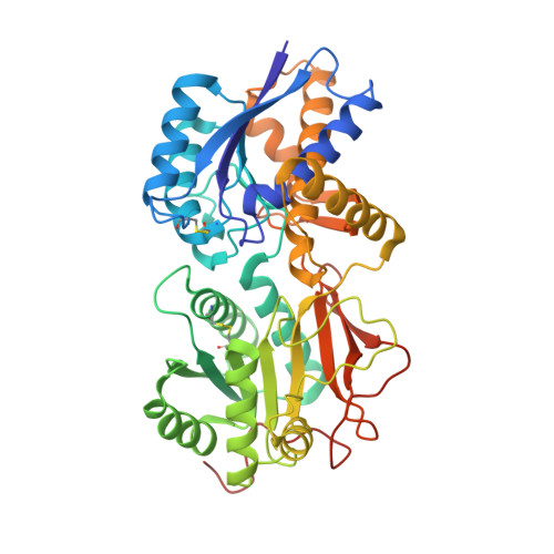
    The binding of atrial natriuretic peptide (ANP) to its receptor requires chloride, and it is chloride concentration dependent. The extracellular domain (ECD) of the ANP receptor (ANPR) contains a chloride near the ANP-binding site, suggesting a possible regulatory role. The bound chloride, however, is completely buried in the polypeptide fold, and its functional role has remained unclear. Here, we have confirmed that chloride is necessary for ANP binding to the recombinant ECD or the full-length ANPR expressed in CHO cells. ECD without chloride (ECD(-)) did not bind ANP. Its binding activity was fully restored by bromide or chloride addition. A new X-ray structure of the bromide-bound ECD is essentially identical to that of the chloride-bound ECD. Furthermore, bromide atoms are localized at the same positions as chloride atoms both in the apo and in the ANP-bound structures, indicating exchangeable and reversible halide binding. Far-UV CD and thermal unfolding data show that ECD(-) largely retains the native structure. Sedimentation equilibrium in the absence of chloride shows that ECD(-) forms a strongly associated dimer, possibly preventing the structural rearrangement of the two monomers that is necessary for ANP binding. The primary and tertiary structures of the chloride-binding site in ANPR are highly conserved among receptor-guanylate cyclases and metabotropic glutamate receptors. The chloride-dependent ANP binding, reversible chloride binding, and the highly conserved chloride-binding site motif suggest a regulatory role for the receptor bound chloride. Chloride-dependent regulation of ANPR may operate in the kidney, modulating ANP-induced natriuresis.


  • Organizational Affiliation

    Department of Biochemistry, University of Nevada School of Medicine, Reno, Nevada 89557, USA.


Macromolecules
Find similar proteins by:  (by identity cutoff)  |  3D Structure
Entity ID: 1
MoleculeChains Sequence LengthOrganismDetailsImage
Atrial natriuretic peptide receptor A
A, B
435Rattus norvegicusMutation(s): 0 
Gene Names: Npr1
EC: 4.6.1.2
UniProt
Find proteins for P18910 (Rattus norvegicus)
Explore P18910 
Go to UniProtKB:  P18910
Entity Groups  
Sequence Clusters30% Identity50% Identity70% Identity90% Identity95% Identity100% Identity
UniProt GroupP18910
Sequence Annotations
Expand
  • Reference Sequence
Oligosaccharides

Help

Entity ID: 2
MoleculeChains Length2D Diagram Glycosylation3D Interactions
2-acetamido-2-deoxy-beta-D-glucopyranose-(1-4)-2-acetamido-2-deoxy-beta-D-glucopyranose
C, D, E
2N-Glycosylation
Glycosylation Resources
GlyTouCan:  G42666HT
GlyCosmos:  G42666HT
GlyGen:  G42666HT
Small Molecules
Ligands 3 Unique
IDChains Name / Formula / InChI Key2D Diagram3D Interactions
NAG
Query on NAG

Download Ideal Coordinates CCD File 
F [auth A],
H [auth A],
M [auth B],
P [auth B]
2-acetamido-2-deoxy-beta-D-glucopyranose
C8 H15 N O6
OVRNDRQMDRJTHS-FMDGEEDCSA-N
SO4
Query on SO4

Download Ideal Coordinates CCD File 
G [auth A]
I [auth A]
J [auth A]
K [auth A]
N [auth B]
G [auth A],
I [auth A],
J [auth A],
K [auth A],
N [auth B],
O [auth B],
Q [auth B],
R [auth B],
S [auth B]
SULFATE ION
O4 S
QAOWNCQODCNURD-UHFFFAOYSA-L
BR
Query on BR

Download Ideal Coordinates CCD File 
L [auth A],
T [auth B]
BROMIDE ION
Br
CPELXLSAUQHCOX-UHFFFAOYSA-M
Experimental Data & Validation

Experimental Data

  • Method: X-RAY DIFFRACTION
  • Resolution: 2.50 Å
  • R-Value Free: 0.243 
  • R-Value Work: 0.205 
  • R-Value Observed: 0.207 
  • Space Group: P 41 21 2
Unit Cell:
Length ( Å )Angle ( ˚ )
a = 120.521α = 90
b = 120.521β = 90
c = 160.613γ = 90
Software Package:
Software NamePurpose
REFMACrefinement
HKL-2000data collection
HKL-2000data reduction
SCALEPACKdata scaling
SOLVEphasing

Structure Validation

View Full Validation Report


Ligand Structure Quality Assessment 


Entry History 

Deposition Data

Revision History  (Full details and data files)

  • Version 1.0: 2010-03-16
    Type: Initial release
  • Version 1.1: 2011-07-13
    Changes: Version format compliance
  • Version 2.0: 2020-07-29
    Type: Remediation
    Reason: Carbohydrate remediation
    Changes: Advisory, Atomic model, Data collection, Derived calculations, Structure summary