3ZTL

Crystal structure of decameric form of Peroxiredoxin I from Schistosoma mansoni


Experimental Data Snapshot

  • Method: X-RAY DIFFRACTION
  • Resolution: 3.00 Å
  • R-Value Free: 0.248 
  • R-Value Work: 0.204 
  • R-Value Observed: 0.206 

wwPDB Validation   3D Report Full Report


This is version 1.1 of the entry. See complete history


Literature

Moonlighting by Different Stressors: Crystal Structure of the Chaperone Species of a 2-Cys Peroxiredoxin.

Saccoccia, F.Di Micco, P.Boumis, G.Brunori, M.Koutris, I.Miele, A.E.Morea, V.Sriratana, P.Williams, D.L.Bellelli, A.Angelucci, F.

(2012) Structure 20: 429

  • DOI: https://doi.org/10.1016/j.str.2012.01.004
  • Primary Citation of Related Structures:  
    3ZTL, 3ZVJ

  • PubMed Abstract: 

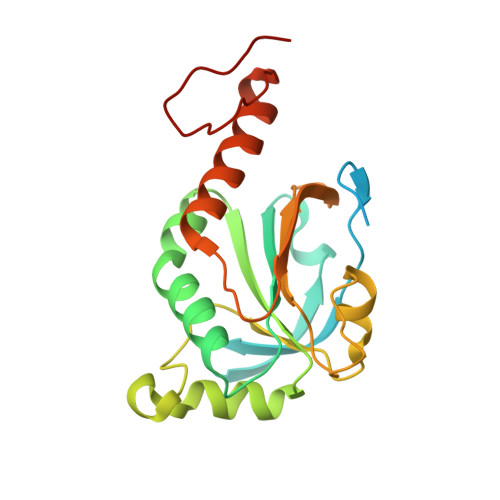
    2-Cys peroxiredoxins (Prxs) play two different roles depending on the physiological status of the cell. They are thioredoxin-dependent peroxidases under low oxidative stress and ATP-independent chaperones upon exposure to high peroxide concentrations. These alternative functions have been associated with changes in the oligomerization state from low-(LMW) to high-molecular-weight (HMW) species. Here we present the structures of Schistosoma mansoni PrxI in both states: the LMW decamer and the HMW 20-mer formed by two stacked decamers. The latter is the structure of a 2-Cys Prx chaperonic form. Comparison of the structures sheds light on the mechanism by which chemical stressors, such as high H(2)O(2) concentration and acidic pH, are sensed and translated into a functional switch in this protein family. We also propose a model to account for the in vivo formation of long filaments of stacked Prx rings.


  • Organizational Affiliation

    Department of Biochemical Sciences, Sapienza University of Rome and Istituto Pasteur-Fondazione Cenci Bolognetti, P.le Aldo Moro 5, 00185 Rome, Italy.


Macromolecules
Find similar proteins by:  (by identity cutoff)  |  3D Structure
Entity ID: 1
MoleculeChains Sequence LengthOrganismDetailsImage
THIOREDOXIN PEROXIDASE
A, B, C, D, E
A, B, C, D, E, F, G, H, I, J
222Schistosoma mansoniMutation(s): 0 
EC: 1.11.1.15
UniProt
Find proteins for O97161 (Schistosoma mansoni)
Explore O97161 
Go to UniProtKB:  O97161
Entity Groups  
Sequence Clusters30% Identity50% Identity70% Identity90% Identity95% Identity100% Identity
UniProt GroupO97161
Sequence Annotations
Expand
  • Reference Sequence
Experimental Data & Validation

Experimental Data

  • Method: X-RAY DIFFRACTION
  • Resolution: 3.00 Å
  • R-Value Free: 0.248 
  • R-Value Work: 0.204 
  • R-Value Observed: 0.206 
  • Space Group: P 1
Unit Cell:
Length ( Å )Angle ( ˚ )
a = 49.507α = 69.12
b = 116.904β = 92
c = 117.349γ = 86.93
Software Package:
Software NamePurpose
PHENIXrefinement
MOSFLMdata reduction
SCALAdata scaling
MOLREPphasing

Structure Validation

View Full Validation Report


Entry History 

Deposition Data

Revision History  (Full details and data files)

  • Version 1.0: 2012-03-21
    Type: Initial release
  • Version 1.1: 2023-12-20
    Changes: Data collection, Database references, Other, Refinement description