3ZS7

Crystal structure of pyridoxal kinase from Trypanosoma brucei


Experimental Data Snapshot

  • Method: X-RAY DIFFRACTION
  • Resolution: 2.00 Å
  • R-Value Free: 0.265 
  • R-Value Work: 0.219 
  • R-Value Observed: 0.222 

wwPDB Validation   3D Report Full Report


Ligand Structure Quality Assessment 


This is version 1.3 of the entry. See complete history


Literature

Chemical, Genetic and Structural Assessment of Pyridoxal Kinase as a Drug Target in the African Trypanosome.

Jones, D.C.Alphey, M.S.Wyllie, S.Fairlamb, A.H.

(2012) Mol Microbiol 86: 51

  • DOI: https://doi.org/10.1111/j.1365-2958.2012.08189.x
  • Primary Citation of Related Structures:  
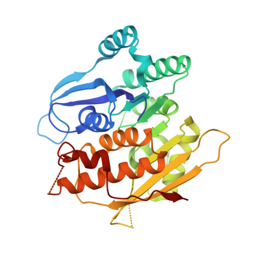
    3ZS7

  • PubMed Abstract: 

    Pyridoxal-5'-phosphate (vitamin B(6) ) is an essential cofactor for many important enzymatic reactions such as transamination and decarboxylation. African trypanosomes are unable to synthesise vitamin B(6) de novo and rely on uptake of B(6) vitamers such as pyridoxal and pyridoxamine from their hosts, which are subsequently phosphorylated by pyridoxal kinase (PdxK). A conditional null mutant of PdxK was generated in Trypanosoma brucei bloodstream forms showing that this enzyme is essential for growth of the parasite in vitro and for infectivity in mice. Activity of recombinant T. brucei PdxK was comparable to previously published work having a specific activity of 327 ± 13 mU mg(-1) and a K(m)(app) with respect to pyridoxal of 29.6 ± 3.9 µM. A coupled assay was developed demonstrating that the enzyme has equivalent catalytic efficiency with pyridoxal, pyridoxamine and pyridoxine, and that ginkgotoxin is an effective pseudo substrate. A high resolution structure of PdxK in complex with ATP revealed important structural differences with the human enzyme. These findings suggest that pyridoxal kinase is an essential and druggable target that could lead to much needed alternative treatments for this devastating disease.


  • Organizational Affiliation

    Division of Biological Chemistry & Drug Discovery, College of Life Sciences, University of Dundee, Dundee, UK.


Macromolecules
Find similar proteins by:  (by identity cutoff)  |  3D Structure
Entity ID: 1
MoleculeChains Sequence LengthOrganismDetailsImage
PYRIDOXAL KINASE300Trypanosoma brucei brucei TREU927Mutation(s): 0 
EC: 2.7.1.35
UniProt
Find proteins for O15927 (Trypanosoma brucei)
Explore O15927 
Go to UniProtKB:  O15927
Entity Groups  
Sequence Clusters30% Identity50% Identity70% Identity90% Identity95% Identity100% Identity
UniProt GroupO15927
Sequence Annotations
Expand
  • Reference Sequence
Small Molecules
Ligands 2 Unique
IDChains Name / Formula / InChI Key2D Diagram3D Interactions
ATP
Query on ATP

Download Ideal Coordinates CCD File 
C [auth A]ADENOSINE-5'-TRIPHOSPHATE
C10 H16 N5 O13 P3
ZKHQWZAMYRWXGA-KQYNXXCUSA-N
PO4
Query on PO4

Download Ideal Coordinates CCD File 
B [auth A]PHOSPHATE ION
O4 P
NBIIXXVUZAFLBC-UHFFFAOYSA-K
Experimental Data & Validation

Experimental Data

  • Method: X-RAY DIFFRACTION
  • Resolution: 2.00 Å
  • R-Value Free: 0.265 
  • R-Value Work: 0.219 
  • R-Value Observed: 0.222 
  • Space Group: C 2 2 21
Unit Cell:
Length ( Å )Angle ( ˚ )
a = 69.21α = 90
b = 80β = 90
c = 106.39γ = 90
Software Package:
Software NamePurpose
REFMACrefinement
XDSdata reduction
XSCALEdata scaling
MOLREPphasing

Structure Validation

View Full Validation Report


Ligand Structure Quality Assessment 


Entry History 

Deposition Data

Revision History  (Full details and data files)

  • Version 1.0: 2012-06-20
    Type: Initial release
  • Version 1.1: 2012-10-03
    Changes: Database references
  • Version 1.2: 2017-06-28
    Changes: Data collection
  • Version 1.3: 2023-12-20
    Changes: Data collection, Database references, Derived calculations, Other, Refinement description