3ZGV

Structure of human SIRT2 in complex with ADP-ribose


Experimental Data Snapshot

  • Method: X-RAY DIFFRACTION
  • Resolution: 2.27 Å
  • R-Value Free: 0.188 
  • R-Value Work: 0.149 
  • R-Value Observed: 0.154 

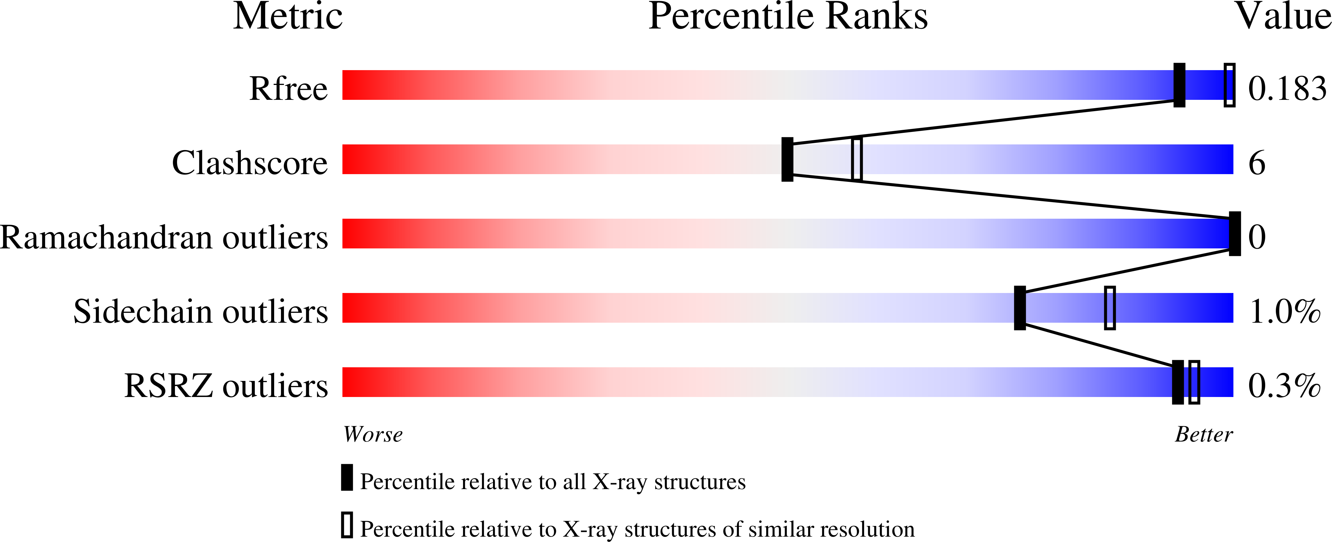
wwPDB Validation   3D Report Full Report


This is version 1.3 of the entry. See complete history


Literature

Crystal Structure Analysis of Human Sirt2 and its Adp-Ribose Complex

Moniot, S.Schutkowski, M.Steegborn, C.

(2013) J Struct Biol 182: 136

  • DOI: https://doi.org/10.1016/j.jsb.2013.02.012
  • Primary Citation of Related Structures:  
    3ZGO, 3ZGV

  • PubMed Abstract: 

    Sirtuins are NAD(+)-dependent protein deacetylases that regulate metabolism and aging-related processes. Sirt2 is the only cytoplasmic isoform among the seven mamalian Sirtuins (Sirt1-7) and structural information concerning this isoform is limited. We crystallized Sirt2 in complex with a product analog, ADP-ribose, and solved this first crystal structure of a Sirt2 ligand complex at 2.3Å resolution. Additionally, we re-refined the structure of the Sirt2 apoform and analyzed the conformational changes associated with ligand binding to derive insights into the dynamics of the enzyme. Our analyses also provide information on Sirt2 peptide substrate binding and structural states of a Sirt2-specific protein region, and our insights and the novel Sirt2 crystal form provide helpful tools for the development of Sirt2 specific inhibitors.


  • Organizational Affiliation

    Department of Biochemistry, University of Bayreuth, 95440 Bayreuth, Germany.


Macromolecules
Find similar proteins by:  (by identity cutoff)  |  3D Structure
Entity ID: 1
MoleculeChains Sequence LengthOrganismDetailsImage
NAD-DEPENDENT PROTEIN DEACETYLASE SIRTUIN-2
A, B
325Homo sapiensMutation(s): 0 
EC: 3.5.1
UniProt & NIH Common Fund Data Resources
Find proteins for Q8IXJ6 (Homo sapiens)
Explore Q8IXJ6 
Go to UniProtKB:  Q8IXJ6
PHAROS:  Q8IXJ6
GTEx:  ENSG00000068903 
Entity Groups  
Sequence Clusters30% Identity50% Identity70% Identity90% Identity95% Identity100% Identity
UniProt GroupQ8IXJ6
Sequence Annotations
Expand
  • Reference Sequence
Small Molecules
Ligands 5 Unique
IDChains Name / Formula / InChI Key2D Diagram3D Interactions
AR6
Query on AR6

Download Ideal Coordinates CCD File 
E [auth A],
K [auth B]
[(2R,3S,4R,5R)-5-(6-AMINOPURIN-9-YL)-3,4-DIHYDROXY-OXOLAN-2-YL]METHYL [HYDROXY-[[(2R,3S,4R,5S)-3,4,5-TRIHYDROXYOXOLAN-2-YL]METHOXY]PHOSPHORYL] HYDROGEN PHOSPHATE
C15 H23 N5 O14 P2
SRNWOUGRCWSEMX-ZQSHOCFMSA-N
PGE
Query on PGE

Download Ideal Coordinates CCD File 
N [auth B]TRIETHYLENE GLYCOL
C6 H14 O4
ZIBGPFATKBEMQZ-UHFFFAOYSA-N
GOL
Query on GOL

Download Ideal Coordinates CCD File 
M [auth B]GLYCEROL
C3 H8 O3
PEDCQBHIVMGVHV-UHFFFAOYSA-N
ZN
Query on ZN

Download Ideal Coordinates CCD File 
C [auth A],
I [auth B]
ZINC ION
Zn
PTFCDOFLOPIGGS-UHFFFAOYSA-N
ACT
Query on ACT

Download Ideal Coordinates CCD File 
D [auth A]
F [auth A]
G [auth A]
H [auth A]
J [auth B]
D [auth A],
F [auth A],
G [auth A],
H [auth A],
J [auth B],
L [auth B]
ACETATE ION
C2 H3 O2
QTBSBXVTEAMEQO-UHFFFAOYSA-M
Experimental Data & Validation

Experimental Data

  • Method: X-RAY DIFFRACTION
  • Resolution: 2.27 Å
  • R-Value Free: 0.188 
  • R-Value Work: 0.149 
  • R-Value Observed: 0.154 
  • Space Group: P 21 21 21
Unit Cell:
Length ( Å )Angle ( ˚ )
a = 77.86α = 90
b = 77.98β = 90
c = 114.33γ = 90
Software Package:
Software NamePurpose
PHENIXrefinement
XDSdata reduction
XSCALEdata scaling
MOLREPphasing

Structure Validation

View Full Validation Report


Entry History 

Deposition Data

Revision History  (Full details and data files)

  • Version 1.0: 2013-03-13
    Type: Initial release
  • Version 1.1: 2013-04-03
    Changes: Database references
  • Version 1.2: 2013-05-08
    Changes: Database references
  • Version 1.3: 2023-12-20
    Changes: Data collection, Database references, Derived calculations, Other, Refinement description, Structure summary