Structural insights into alginate binding by bacterial cell-surface protein
Temtrirath, K., Murata, K., Hashimoto, W.(2014) Carbohydr Res 404C: 39-45
- PubMed: 25665777
- DOI: https://doi.org/10.1016/j.carres.2014.11.008
- Primary Citation of Related Structures:
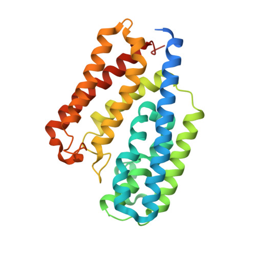
3WSC - PubMed Abstract:
A gram-negative Sphingomonas sp. strain A1 inducibly forms a mouth-like pit on the cell surface in the presence of alginate and directly incorporates polymers into the cytoplasm via the pit and ABC transporter. Among the bacterial proteins involved in import of alginate, a cell-surface EfeO-like Algp7 shows an ability to bind alginate, suggesting its contribution to accumulate alginate in the pit. Here, we show identification of its positively charged cluster involved in alginate binding using X-ray crystallography, docking simulation, and site-directed mutagenesis. The tertiary structure of Algp7 was determined at a high resolution (1.99Å) by molecular replacement, although no alginates were included in the structure. Thus, an in silico model of Algp7/oligoalginate was constructed by docking simulation using atomic coordinates of Algp7 and alginate oligosaccharides, where some charged residues were found to be potential candidates for alginate binding. Site-directed mutagenesis was conducted and five purified mutants K68A, K69A, E194A, N221A, and K68A/K69A were subjected to a binding assay. UV absorption difference spectroscopy along with differential scanning fluorimetry analysis indicated that K68A/K69A exhibited a significant reduction in binding affinity with alginate than wild-type Algp7. Based on these data, Lys68/Lys69 residues of Algp7 probably play an important role in binding alginate.
Organizational Affiliation:
Laboratory of Basic and Applied Molecular Biotechnology, Division of Food Science and Biotechnology, Graduate School of Agriculture, Kyoto University, Gokasho, Uji, Kyoto 611-0011, Japan.