3WEU

Crystal structure of the L-Lys epsilon-oxidase from Marinomonas mediterranea


Experimental Data Snapshot

  • Method: X-RAY DIFFRACTION
  • Resolution: 1.93 Å
  • R-Value Free: 0.178 
  • R-Value Work: 0.143 
  • R-Value Observed: 0.143 

wwPDB Validation   3D Report Full Report


This is version 1.0 of the entry. See complete history


Literature

X-Ray crystallographic evidence for the presence of the cysteine tryptophylquinone cofactor in L-lysine {varepsilon}-oxidase from Marinomonas mediterranea

Okazaki, S.Nakano, S.Matsui, D.Akaji, S.Inagaki, K.Asano, Y.

(2013) J Biochem 154: 233-236

  • DOI: https://doi.org/10.1093/jb/mvt070
  • Primary Citation of Related Structures:  
    3WEU, 3WEV

  • PubMed Abstract: 

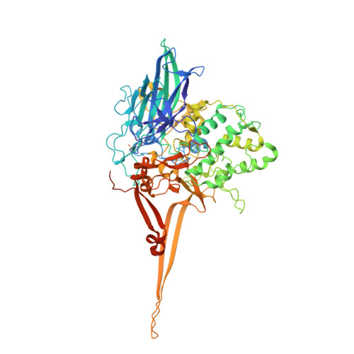
    We have determined the x-ray crystal structure of L-lysine ε-oxidase from Marinomonas mediterranea in its native and L-lysine-complex forms at 1.94- and 1.99-Å resolution, respectively. In the native enzyme, electron densities clearly indicate the presence of cysteine tryptophylquinone (CTQ) previously identified in quinohemoprotein amine dehydrogenase. In the L-lysine-complex, an electron density corresponding to the bound L-lysine shows that its ε-amino group is attached to the C6 carbonyl group of CTQ, suggesting the formation of a Schiff-base intermediate. Collectively, the present crystal structure provides the first example of an enzyme employing a tryptophylquinone cofactor in an amine oxidase.


  • Organizational Affiliation

    Biotechnology Research Center and Department of Biotechnology, Toyama Prefectural University, 5180 Kurokawa, Imizu, Toyama 939-0398, Japan.


Macromolecules
Find similar proteins by:  (by identity cutoff)  |  3D Structure
Entity ID: 1
MoleculeChains Sequence LengthOrganismDetailsImage
L-lysine 6-oxidase
A, B
726Marinomonas mediterranea MMB-1Mutation(s): 0 
Gene Names: lodA
EC: 1.4.3.20
UniProt
Find proteins for F2JXJ3 (Marinomonas mediterranea (strain ATCC 700492 / JCM 21426 / NBRC 103028 / MMB-1))
Explore F2JXJ3 
Go to UniProtKB:  F2JXJ3
Entity Groups  
Sequence Clusters30% Identity50% Identity70% Identity90% Identity95% Identity100% Identity
UniProt GroupF2JXJ3
Sequence Annotations
Expand
  • Reference Sequence
Small Molecules
Ligands 3 Unique
IDChains Name / Formula / InChI Key2D Diagram3D Interactions
SO4
Query on SO4

Download Ideal Coordinates CCD File 
C [auth A]
D [auth A]
E [auth A]
P [auth B]
Q [auth B]
C [auth A],
D [auth A],
E [auth A],
P [auth B],
Q [auth B],
R [auth B]
SULFATE ION
O4 S
QAOWNCQODCNURD-UHFFFAOYSA-L
DIO
Query on DIO

Download Ideal Coordinates CCD File 
F [auth A],
S [auth B]
1,4-DIETHYLENE DIOXIDE
C4 H8 O2
RYHBNJHYFVUHQT-UHFFFAOYSA-N
EDO
Query on EDO

Download Ideal Coordinates CCD File 
AA [auth B]
BA [auth B]
G [auth A]
H [auth A]
I [auth A]
AA [auth B],
BA [auth B],
G [auth A],
H [auth A],
I [auth A],
J [auth A],
K [auth A],
L [auth A],
M [auth A],
N [auth A],
O [auth A],
T [auth B],
U [auth B],
V [auth B],
W [auth B],
X [auth B],
Y [auth B],
Z [auth B]
1,2-ETHANEDIOL
C2 H6 O2
LYCAIKOWRPUZTN-UHFFFAOYSA-N
Modified Residues  2 Unique
IDChains TypeFormula2D DiagramParent
CSD
Query on CSD
A, B
L-PEPTIDE LINKINGC3 H7 N O4 SCYS
TRQ
Query on TRQ
A, B
L-PEPTIDE LINKINGC11 H10 N2 O4TRP
Experimental Data & Validation

Experimental Data

  • Method: X-RAY DIFFRACTION
  • Resolution: 1.93 Å
  • R-Value Free: 0.178 
  • R-Value Work: 0.143 
  • R-Value Observed: 0.143 
  • Space Group: C 1 2 1
Unit Cell:
Length ( Å )Angle ( ˚ )
a = 194.089α = 90
b = 125.746β = 99.22
c = 70.591γ = 90
Software Package:
Software NamePurpose
HKL-2000data collection
PHENIXmodel building
REFMACrefinement
HKL-2000data reduction
HKL-2000data scaling
PHENIXphasing

Structure Validation

View Full Validation Report


Entry History 

Deposition Data

Revision History  (Full details and data files)

  • Version 1.0: 2013-09-04
    Type: Initial release