


Crystal structure of peptidoglycan hydrolase mutant from Sphingomonas sp. A1
Maruyama, Y., Mikami, B., Hashimoto, W., Murata, K.To be published.
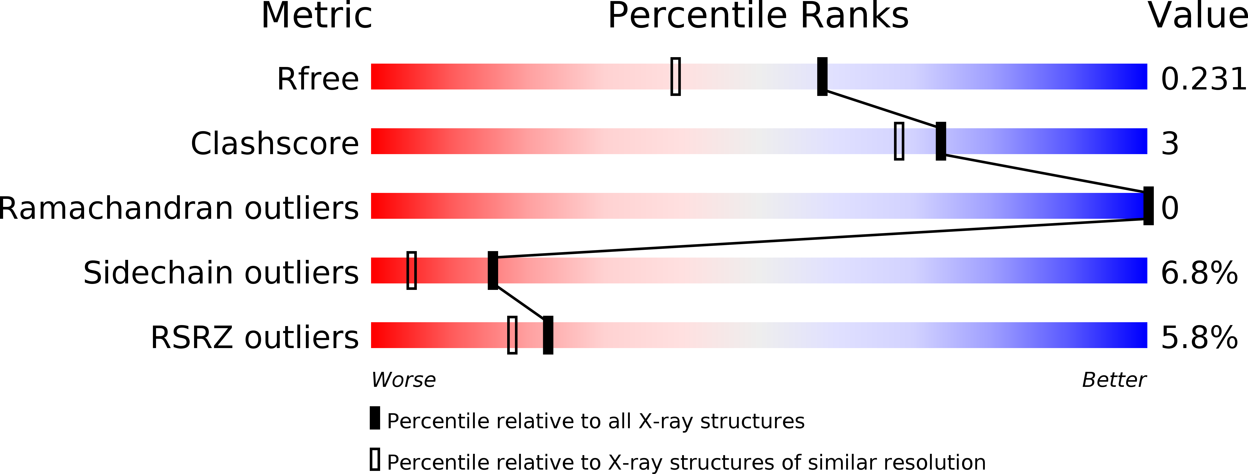
Experimental Data Snapshot
wwPDB Validation 3D Report Full Report
Entity ID: 1 | |||||
|---|---|---|---|---|---|
| Molecule | Chains | Sequence Length | Organism | Details | Image |
| Peptidoglycan hydrolase FlgJ | 161 | Sphingomonas sp. A1 | Mutation(s): 0 Gene Names: flgJ | 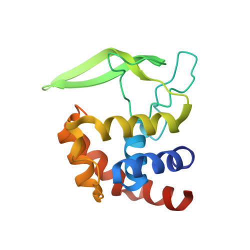 | |
UniProt | |||||
Find proteins for B7XH69 (Sphingomonas sp. A1) Explore B7XH69 Go to UniProtKB: B7XH69 | |||||
Entity Groups | |||||
| Sequence Clusters | 30% Identity50% Identity70% Identity90% Identity95% Identity100% Identity | ||||
| UniProt Group | B7XH69 | ||||
Sequence AnnotationsExpand | |||||
| |||||
| Ligands 1 Unique | |||||
|---|---|---|---|---|---|
| ID | Chains | Name / Formula / InChI Key | 2D Diagram | 3D Interactions | |
| EPE Query on EPE | B [auth A] | 4-(2-HYDROXYETHYL)-1-PIPERAZINE ETHANESULFONIC ACID C8 H18 N2 O4 S JKMHFZQWWAIEOD-UHFFFAOYSA-N |  | ||
| Length ( Å ) | Angle ( ˚ ) |
|---|---|
| a = 48.467 | α = 90 |
| b = 48.467 | β = 90 |
| c = 124.203 | γ = 90 |
| Software Name | Purpose |
|---|---|
| MOLREP | phasing |
| PHENIX | refinement |
| HKL-2000 | data reduction |
| HKL-2000 | data scaling |
RCSB PDB (citation) is hosted by
RCSB PDB is a member of the

