3VDI

Structure of the FMO protein from Pelodictyon phaeum


Experimental Data Snapshot

  • Method: X-RAY DIFFRACTION
  • Resolution: 1.99 Å
  • R-Value Free: 0.190 
  • R-Value Work: 0.162 
  • R-Value Observed: 0.164 

wwPDB Validation   3D Report Full Report


Ligand Structure Quality Assessment 


This is version 1.3 of the entry. See complete history


Literature

Reinterpretation of the electron density at the site of the eighth bacteriochlorophyll in the FMO protein from Pelodictyon phaeum.

Tronrud, D.E.Allen, J.P.

(2012) Photosynth Res 112: 71-74

  • DOI: https://doi.org/10.1007/s11120-012-9735-8
  • Primary Citation of Related Structures:  
    3VDI

  • PubMed Abstract: 

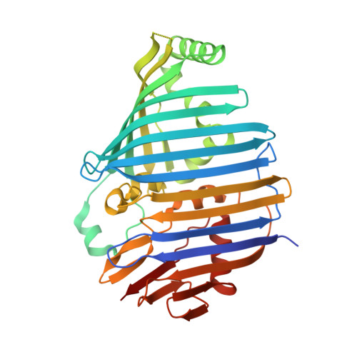
    The Fenna-Matthews-Olson antenna protein from the green bacterium Pelodictyon phaeum mediates the energy transfer from a peripheral antenna complex to the membrane-bound reaction center. The three-dimensional structure of this protein has been previously modeled using X-ray diffraction to a resolution limit of 2.0 Å, with R (work) and R (free) values of 16.6 and 19.9%, respectively (Larson et al., Photosynth Res 107:139-150, 2011). This model shows the protein as consisting of β-sheets surrounding several bacteriochlorophyll cofactors. While most of the model clearly matches the electron density maps, in this paper we re-examine the electron density for a specific feature, namely the eighth bacteriochlorophyll a cofactor. This electron density is now interpreted as arising primarily from the end of an otherwise disordered polyethylene glycol molecule. Additional electron density is present but the density is weak and cannot be unambiguously assigned. The new model has R (work) and R (free) values of 16.2 and 19.0%, respectively.


  • Organizational Affiliation

    Department of Biochemistry and Biophysics, Oregon State University, Corvallis, OR 97331, USA. tronrudd@science.oregonstate.edu


Macromolecules
Find similar proteins by:  (by identity cutoff)  |  3D Structure
Entity ID: 1
MoleculeChains Sequence LengthOrganismDetailsImage
bacteriochlorophyll A protein367Pelodictyon phaeumMutation(s): 0 
Entity Groups  
Sequence Clusters30% Identity50% Identity70% Identity90% Identity95% Identity100% Identity
Sequence Annotations
Expand
  • Reference Sequence
Small Molecules
Ligands 2 Unique
IDChains Name / Formula / InChI Key2D Diagram3D Interactions
BCL
Query on BCL

Download Ideal Coordinates CCD File 
B [auth A]
C [auth A]
D [auth A]
E [auth A]
F [auth A]
B [auth A],
C [auth A],
D [auth A],
E [auth A],
F [auth A],
G [auth A],
H [auth A]
BACTERIOCHLOROPHYLL A
C55 H74 Mg N4 O6
DSJXIQQMORJERS-AGGZHOMASA-M
PG4
Query on PG4

Download Ideal Coordinates CCD File 
I [auth A],
J [auth A],
K [auth A],
L [auth A]
TETRAETHYLENE GLYCOL
C8 H18 O5
UWHCKJMYHZGTIT-UHFFFAOYSA-N
Experimental Data & Validation

Experimental Data

  • Method: X-RAY DIFFRACTION
  • Resolution: 1.99 Å
  • R-Value Free: 0.190 
  • R-Value Work: 0.162 
  • R-Value Observed: 0.164 
  • Space Group: P 63
Unit Cell:
Length ( Å )Angle ( ˚ )
a = 83.967α = 90
b = 83.967β = 90
c = 115.724γ = 120
Software Package:
Software NamePurpose
CBASSdata collection
PHENIXmodel building
BUSTERrefinement
MOSFLMdata reduction
SCALAdata scaling
PHENIXphasing

Structure Validation

View Full Validation Report


Ligand Structure Quality Assessment 


Entry History 

Deposition Data

Revision History  (Full details and data files)

  • Version 1.0: 2012-01-25
    Type: Initial release
  • Version 1.1: 2012-04-11
    Changes: Database references
  • Version 1.2: 2012-05-09
    Changes: Database references
  • Version 1.3: 2023-09-13
    Changes: Data collection, Database references, Derived calculations, Refinement description