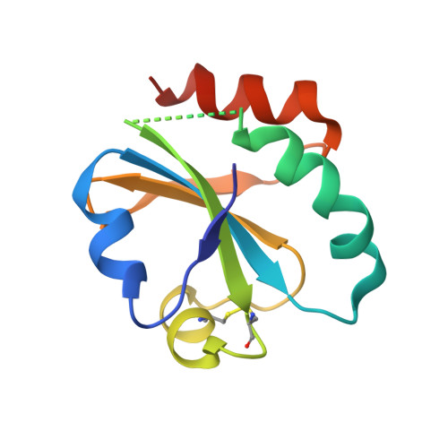
3UJ1

Crystal structure of the third thioredoxin domain of human ERp46


Experimental Data Snapshot

  • Method: X-RAY DIFFRACTION
  • Resolution: 2.65 Å
  • R-Value Free: 0.259 
  • R-Value Work: 0.229 
  • R-Value Observed: 0.230 

wwPDB Validation   3D Report Full Report


This is version 1.3 of the entry. See complete history


Literature

Peptide Binding by Catalytic Domains of the Protein Disulfide Isomerase-Related Protein ERp46.

Funkner, A.Parthier, C.Schutkowski, M.Zerweck, J.Lilie, H.Gyrych, N.Fischer, G.Stubbs, M.T.Ferrari, D.M.

(2013) J Mol Biol 425: 1340-1362

  • DOI: https://doi.org/10.1016/j.jmb.2013.01.029
  • Primary Citation of Related Structures:  
    3UJ1

  • PubMed Abstract: 

    The protein disulfide isomerase (PDI) family member ERp46/endoPDI/thioredoxin domain-containing protein 5 is preferentially expressed in a limited number of tissues, where it may function as a survival factor for nitrosative stress in vivo. It is involved in insulin production as well as in adiponectin signaling and interacts specifically with the redox-regulatory endoplasmic reticulum proteins endoplasmic oxidoreductin 1α (Ero1α) and peroxiredoxin-4. Here, we show that ERp46, although lacking a PDI-like redox-inactive b'-thioredoxin domain with its hydrophobic substrate binding site, is able to bind to a large pool of peptides containing aromatic and basic residues via all three of its catalytic domains (a(0), a and a'), though the a(0) domain may contain the primary binding site. ERp46, which shows relatively higher activity as a disulfide-reductase than as an oxidase/isomerase in vitro compared to PDI and ERp57, possesses chaperone activity in vivo, a property also shared by the C-terminal a' domain. A crystal structure of the a' domain is also presented, offering a view of possible substrate binding sites within catalytic domains of PDI proteins.


  • Organizational Affiliation

    Max Planck Research Unit for Enzymology of Protein Folding, Weinbergweg 22, 06120 Halle (Saale), Germany.


Macromolecules
Find similar proteins by:  (by identity cutoff)  |  3D Structure
Entity ID: 1
MoleculeChains Sequence LengthOrganismDetailsImage
Thioredoxin domain-containing protein 5110Homo sapiensMutation(s): 0 
Gene Names: TLP46TXNDC5UNQ364/PRO700
UniProt & NIH Common Fund Data Resources
Find proteins for Q8NBS9 (Homo sapiens)
Explore Q8NBS9 
Go to UniProtKB:  Q8NBS9
PHAROS:  Q8NBS9
GTEx:  ENSG00000239264 
Entity Groups  
Sequence Clusters30% Identity50% Identity70% Identity90% Identity95% Identity100% Identity
UniProt GroupQ8NBS9
Sequence Annotations
Expand
  • Reference Sequence
Experimental Data & Validation

Experimental Data

  • Method: X-RAY DIFFRACTION
  • Resolution: 2.65 Å
  • R-Value Free: 0.259 
  • R-Value Work: 0.229 
  • R-Value Observed: 0.230 
  • Space Group: P 32 2 1
Unit Cell:
Length ( Å )Angle ( ˚ )
a = 63.58α = 90
b = 63.58β = 90
c = 68.68γ = 120
Software Package:
Software NamePurpose
MOSFLMdata reduction
SCALAdata scaling
PHASERphasing
REFMACrefinement
PDB_EXTRACTdata extraction
AUTOMARdata collection

Structure Validation

View Full Validation Report


Entry History 

Deposition Data

Revision History  (Full details and data files)

  • Version 1.0: 2012-11-07
    Type: Initial release
  • Version 1.1: 2013-03-06
    Changes: Database references
  • Version 1.2: 2013-04-17
    Changes: Database references
  • Version 1.3: 2023-09-13
    Changes: Data collection, Database references, Refinement description