Crystal structure of the MrkD1P receptor binding domain of Klebsiella pneumoniae and identification of the human collagen V binding interface.
Rego, A.T., Johnson, J.G., Geibel, S., Enguita, F.J., Clegg, S., Waksman, G.(2012) Mol Microbiol 86: 882-893
- PubMed: 22988966
- DOI: https://doi.org/10.1111/mmi.12023
- Primary Citation of Related Structures:
3U4K - PubMed Abstract:
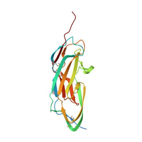
Klebsiella species are members of the family enterobacteriaceae, opportunistic pathogens that are among the eight most prevalent infectious agents in hospitals. Among other virulence factors in Klebsiella, type 3 pili exhibit a unique binding pattern in the human kidney via interaction of two MrkD adhesion variants 1C1 and 1P to type IV and/or V collagen. However, very little is known about the nature of this recognition. Here we present the crystal structure of the plasmid born MrkD1P receptor domain (MrkDrd). The structure reveals a jelly-roll β-barrel fold comprising 17 β-strands very similar to the receptor domain of GafD, the tip adhesin from the F17 pilus that recognizes n-acetyl-d-glucosamine (GlcNAc). Analysis of collagen V binding of different MrkD1P mutants revealed that two regions were responsible for its binding: a pocket, that aligns approximately with the GlcNAc binding pocket of GafD involving residues R105 and Y155, and a transversally oriented patch that spans strands β2a, β9b and β6 including residues V49, T52, V91, R102 and I136. Taken together, these data provide structural and functional insights on MrkD1P recognition of host cells, providing a tool for future development of rationally designed drugs with the prospect of blocking Klebsiella adhesion to collagen V.
Organizational Affiliation:
Institute of Structural and Molecular Biology, University College London and Birkbeck College, Malet Street, London, WC1E 7HX, UK.