3U2L

Crystal structure of human ALR mutant C142S.


Experimental Data Snapshot

  • Method: X-RAY DIFFRACTION
  • Resolution: 1.95 Å
  • R-Value Free: 0.265 
  • R-Value Work: 0.189 
  • R-Value Observed: 0.196 

wwPDB Validation   3D Report Full Report


Ligand Structure Quality Assessment 


This is version 1.2 of the entry. See complete history


Literature

An electron-transfer path through an extended disulfide relay system: the case of the redox protein ALR.

Banci, L.Bertini, I.Calderone, V.Cefaro, C.Ciofi-Baffoni, S.Gallo, A.Tokatlidis, K.

(2012) J Am Chem Soc 134: 1442-1445

  • DOI: https://doi.org/10.1021/ja209881f
  • Primary Citation of Related Structures:  
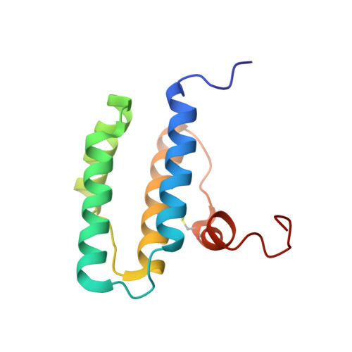
    3U2L, 3U2M

  • PubMed Abstract: 

    The oxidative folding mechanism in the intermembrane space of human mitochondria underpins a disulfide relay system consisting of the import receptor Mia40 and the homodimeric FAD-dependent thiol oxidase ALR. The flavoprotein ALR receives two electrons per subunit from Mia40, which are then donated through one-electron reactions to two cytochrome c molecules, thus mediating a switch from two-electron to one-electron transfer. We dissect here the mechanism of the electron flux within ALR, characterizing at the atomic level the ALR intermediates that allow electrons to rapidly flow to cytochrome c. The intermediate critical for the electron-transfer process implies the formation of a specific inter-subunit disulfide which exclusively allows electron flow from Mia40 to FAD. This finding allows us to present a complete model for the electron-transfer pathway in ALR.


  • Organizational Affiliation

    Magnetic Resonance Center, University of Florence, via L. Sacconi 6, Sesto Fiorentino, Italy. banci@cerm.unifi.it


Macromolecules
Find similar proteins by:  (by identity cutoff)  |  3D Structure
Entity ID: 1
MoleculeChains Sequence LengthOrganismDetailsImage
FAD-linked sulfhydryl oxidase ALR115Homo sapiensMutation(s): 3 
Gene Names: GFERALRHERV1HPO
EC: 1.8.3.2
UniProt & NIH Common Fund Data Resources
Find proteins for P55789 (Homo sapiens)
Explore P55789 
Go to UniProtKB:  P55789
PHAROS:  P55789
GTEx:  ENSG00000127554 
Entity Groups  
Sequence Clusters30% Identity50% Identity70% Identity90% Identity95% Identity100% Identity
UniProt GroupP55789
Sequence Annotations
Expand
  • Reference Sequence
Small Molecules
Ligands 1 Unique
IDChains Name / Formula / InChI Key2D Diagram3D Interactions
FAD
Query on FAD

Download Ideal Coordinates CCD File 
B [auth A]FLAVIN-ADENINE DINUCLEOTIDE
C27 H33 N9 O15 P2
VWWQXMAJTJZDQX-UYBVJOGSSA-N
Experimental Data & Validation

Experimental Data

  • Method: X-RAY DIFFRACTION
  • Resolution: 1.95 Å
  • R-Value Free: 0.265 
  • R-Value Work: 0.189 
  • R-Value Observed: 0.196 
  • Space Group: C 2 2 21
Unit Cell:
Length ( Å )Angle ( ˚ )
a = 50.894α = 90
b = 76.641β = 90
c = 62.56γ = 90
Software Package:
Software NamePurpose
CrysalisProdata collection
MOLREPphasing
REFMACrefinement
MOSFLMdata reduction
SCALAdata scaling

Structure Validation

View Full Validation Report


Ligand Structure Quality Assessment 


Entry History 

Deposition Data

Revision History  (Full details and data files)

  • Version 1.0: 2012-06-06
    Type: Initial release
  • Version 1.1: 2017-11-08
    Changes: Refinement description
  • Version 1.2: 2023-09-13
    Changes: Data collection, Database references, Derived calculations, Refinement description