


Crystal structure of peptide deformylase from ehrlichia chaffeensis in complex with actinonin
Seattle Structural Genomics Center for Infectious Disease (SSGCID), Abendroth, J., Clifton, M.C., Edwards, T.E., Staker, B.L.To be published.
Experimental Data Snapshot
Entity ID: 1 | |||||
|---|---|---|---|---|---|
| Molecule | Chains | Sequence Length | Organism | Details | Image |
| Peptide deformylase 1 | 190 | Ehrlichia chaffeensis str. Arkansas | Mutation(s): 0 Gene Names: def, def1, ECH_0073 EC: 3.5.1.88 | 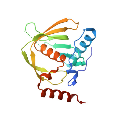 | |
UniProt | |||||
Find proteins for Q2GI30 (Ehrlichia chaffeensis (strain ATCC CRL-10679 / Arkansas)) Explore Q2GI30 Go to UniProtKB: Q2GI30 | |||||
Entity Groups | |||||
| Sequence Clusters | 30% Identity50% Identity70% Identity90% Identity95% Identity100% Identity | ||||
| UniProt Group | Q2GI30 | ||||
Sequence AnnotationsExpand | |||||
| |||||
| Ligands 3 Unique | |||||
|---|---|---|---|---|---|
| ID | Chains | Name / Formula / InChI Key | 2D Diagram | 3D Interactions | |
| BB2 Query on BB2 | E [auth A] | ACTINONIN C19 H35 N3 O5 XJLATMLVMSFZBN-VYDXJSESSA-N |  | ||
| ZN Query on ZN | B [auth A] | ZINC ION Zn PTFCDOFLOPIGGS-UHFFFAOYSA-N |  | ||
| CL Query on CL | C [auth A], D [auth A] | CHLORIDE ION Cl VEXZGXHMUGYJMC-UHFFFAOYSA-M |  | ||
| Length ( Å ) | Angle ( ˚ ) |
|---|---|
| a = 83.89 | α = 90 |
| b = 33.02 | β = 91.17 |
| c = 68.14 | γ = 90 |
| Software Name | Purpose |
|---|---|
| XDS | data scaling |
| PHASER | phasing |
| REFMAC | refinement |
RCSB PDB (citation) is hosted by
RCSB PDB is a member of the

