3TTO

Crystal structure of Leuconostoc mesenteroides NRRL B-1299 N-terminally truncated dextransucrase DSR-E in triclinic form


Experimental Data Snapshot

  • Method: X-RAY DIFFRACTION
  • Resolution: 3.30 Å
  • R-Value Free: 0.291 
  • R-Value Work: 0.224 
  • R-Value Observed: 0.227 

wwPDB Validation   3D Report Full Report


This is version 1.3 of the entry. See complete history


Literature

Functional and structural characterization of alpha-(1-2) branching sucrase derived from DSR-E glucansucrase

Brison, Y.Pijning, T.Malbert, Y.Fabre, E.Mourey, L.Morel, S.Potocki-Veronese, G.Monsan, P.Tranier, S.Remaud-Simeon, M.Dijkstra, B.W.

(2012) J Biol Chem 287: 7915-7924

  • DOI: https://doi.org/10.1074/jbc.M111.305078
  • Primary Citation of Related Structures:  
    3TTO, 3TTQ

  • PubMed Abstract: 

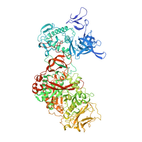
    ΔN(123)-glucan-binding domain-catalytic domain 2 (ΔN(123)-GBD-CD2) is a truncated form of the bifunctional glucansucrase DSR-E from Leuconostoc mesenteroides NRRL B-1299. It was constructed by rational truncation of GBD-CD2, which harbors the second catalytic domain of DSR-E. Like GBD-CD2, this variant displays α-(1→2) branching activity when incubated with sucrose as glucosyl donor and (oligo-)dextran as acceptor, transferring glucosyl residues to the acceptor via a ping-pong bi-bi mechanism. This allows the formation of prebiotic molecules containing controlled amounts of α-(1→2) linkages. The crystal structure of the apo α-(1→2) branching sucrase ΔN(123)-GBD-CD2 was solved at 1.90 Å resolution. The protein adopts the unusual U-shape fold organized in five distinct domains, also found in GTF180-ΔN and GTF-SI glucansucrases of glycoside hydrolase family 70. Residues forming subsite -1, involved in binding the glucosyl residue of sucrose and catalysis, are strictly conserved in both GTF180-ΔN and ΔN(123)-GBD-CD2. Subsite +1 analysis revealed three residues (Ala-2249, Gly-2250, and Phe-2214) that are specific to ΔN(123)-GBD-CD2. Mutation of these residues to the corresponding residues found in GTF180-ΔN showed that Ala-2249 and Gly-2250 are not directly involved in substrate binding and regiospecificity. In contrast, mutant F2214N had lost its ability to branch dextran, although it was still active on sucrose alone. Furthermore, three loops belonging to domains A and B at the upper part of the catalytic gorge are also specific to ΔN(123)-GBD-CD2. These distinguishing features are also proposed to be involved in the correct positioning of dextran acceptor molecules allowing the formation of α-(1→2) branches.


  • Organizational Affiliation

    Université de Toulouse, INSA, UPS, INP, LISBP, F-31077 Toulouse, France.


Macromolecules
Find similar proteins by:  (by identity cutoff)  |  3D Structure
Entity ID: 1
MoleculeChains Sequence LengthOrganismDetailsImage
Dextransucrase
A, B, C, D
1,108Leuconostoc mesenteroidesMutation(s): 0 
Gene Names: dsr-EdsrE
EC: 2.4.1.5
UniProt
Find proteins for Q8G9Q2 (Leuconostoc mesenteroides)
Explore Q8G9Q2 
Go to UniProtKB:  Q8G9Q2
Entity Groups  
Sequence Clusters30% Identity50% Identity70% Identity90% Identity95% Identity100% Identity
UniProt GroupQ8G9Q2
Sequence Annotations
Expand
  • Reference Sequence
Small Molecules
Ligands 2 Unique
IDChains Name / Formula / InChI Key2D Diagram3D Interactions
GOL
Query on GOL

Download Ideal Coordinates CCD File 
F [auth A]
H [auth B]
I [auth B]
K [auth C]
L [auth C]
F [auth A],
H [auth B],
I [auth B],
K [auth C],
L [auth C],
M [auth C],
O [auth D],
P [auth D],
Q [auth D]
GLYCEROL
C3 H8 O3
PEDCQBHIVMGVHV-UHFFFAOYSA-N
CA
Query on CA

Download Ideal Coordinates CCD File 
E [auth A],
G [auth B],
J [auth C],
N [auth D]
CALCIUM ION
Ca
BHPQYMZQTOCNFJ-UHFFFAOYSA-N
Experimental Data & Validation

Experimental Data

  • Method: X-RAY DIFFRACTION
  • Resolution: 3.30 Å
  • R-Value Free: 0.291 
  • R-Value Work: 0.224 
  • R-Value Observed: 0.227 
  • Space Group: P 1
Unit Cell:
Length ( Å )Angle ( ˚ )
a = 66.84α = 85.36
b = 140.04β = 90.92
c = 155.46γ = 76.85
Software Package:
Software NamePurpose
ADSCdata collection
PHASERphasing
REFMACrefinement
iMOSFLMdata reduction
SCALAdata scaling

Structure Validation

View Full Validation Report


Entry History 

Deposition Data

Revision History  (Full details and data files)

  • Version 1.0: 2012-01-25
    Type: Initial release
  • Version 1.1: 2013-06-26
    Changes: Database references
  • Version 1.2: 2018-01-31
    Changes: Database references
  • Version 1.3: 2023-09-13
    Changes: Data collection, Database references, Derived calculations, Refinement description