3TS4

Human MMP12 in complex with L-glutamate motif inhibitor


Experimental Data Snapshot

  • Method: X-RAY DIFFRACTION
  • Resolution: 1.59 Å
  • R-Value Free: 0.203 
  • R-Value Work: 0.178 
  • R-Value Observed: 0.180 

wwPDB Validation   3D Report Full Report


This is version 1.3 of the entry. See complete history


Literature

Simple pseudo-dipeptides with a P2' glutamate: a novel inhibitor family of matrix metalloproteases and other metzincins.

Devel, L.Beau, F.Amoura, M.Vera, L.Cassar-Lajeunesse, E.Garcia, S.Czarny, B.Stura, E.A.Dive, V.

(2012) J Biol Chem 287: 26647-26656

  • DOI: https://doi.org/10.1074/jbc.M112.380782
  • Primary Citation of Related Structures:  
    3TS4, 3TSK, 3TT4, 3TVC, 4EFS

  • PubMed Abstract: 

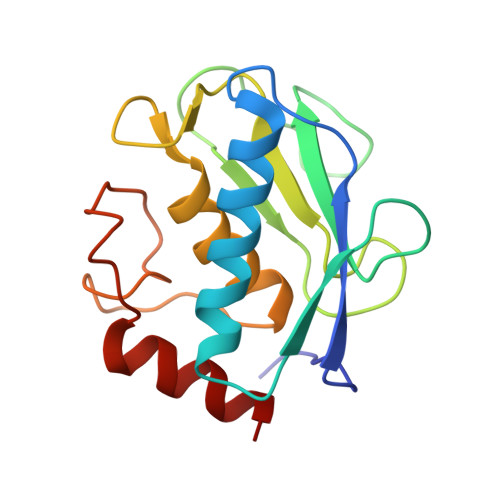
    A series of pseudo-peptides with general formula X-l-Glu-NH(2) (with X corresponding to an acyl moiety with a long aryl-alkyl side chain) have been synthesized, evaluated as inhibitors of matrix metalloproteases (MMPs), and found to display remarkable nanomolar affinity. The loss in potency associated with a substitution of the P(2)' l-glutamate by a l-glutamine corroborates the importance of a carboxylate at this position. The binding mode of some of these inhibitors was characterized in solution and by x-ray crystallography in complex with various MMPs. The x-ray crystal structures reveal an unusual binding mode with the glutamate side chain chelating the active site zinc ion. Competition experiments between these inhibitors and acetohydroxamic acid, a small zinc-binding molecule, are in accord with the crystallographic results. One of these pseudo-dipeptides displays potency and selectivity toward MMP-12 similar to the best MMP-12 inhibitors reported to date. This novel family of pseudo peptides opens new opportunities to develop potent and selective inhibitors for several metzincins.


  • Organizational Affiliation

    CEA (Commissariat à l'Energie Atomique), iBiTec-S, Service d'Ingénierie Moléculaire de Protéines (SIMOPRO), CE Saclay, 91191 Gif/Yvette, Cedex, France. laurent.devel@cea.fr


Macromolecules
Find similar proteins by:  (by identity cutoff)  |  3D Structure
Entity ID: 1
MoleculeChains Sequence LengthOrganismDetailsImage
Macrophage metalloelastase159Homo sapiensMutation(s): 1 
Gene Names: HMEMMP12
EC: 3.4.24.65
UniProt & NIH Common Fund Data Resources
Find proteins for P39900 (Homo sapiens)
Explore P39900 
Go to UniProtKB:  P39900
PHAROS:  P39900
GTEx:  ENSG00000262406 
Entity Groups  
Sequence Clusters30% Identity50% Identity70% Identity90% Identity95% Identity100% Identity
UniProt GroupP39900
Sequence Annotations
Expand
  • Reference Sequence
Small Molecules
Ligands 5 Unique
IDChains Name / Formula / InChI Key2D Diagram3D Interactions
EEG
Query on EEG

Download Ideal Coordinates CCD File 
G [auth A]N-{3-[4-(4-phenylthiophen-2-yl)phenyl]propanoyl}-L-alpha-glutamyl-L-alpha-glutamyl-amide
C29 H31 N3 O7 S
MMIHYNVQXYWTFM-GOTSBHOMSA-N
GOL
Query on GOL

Download Ideal Coordinates CCD File 
I [auth A]GLYCEROL
C3 H8 O3
PEDCQBHIVMGVHV-UHFFFAOYSA-N
IMD
Query on IMD

Download Ideal Coordinates CCD File 
H [auth A]IMIDAZOLE
C3 H5 N2
RAXXELZNTBOGNW-UHFFFAOYSA-O
ZN
Query on ZN

Download Ideal Coordinates CCD File 
B [auth A],
C [auth A]
ZINC ION
Zn
PTFCDOFLOPIGGS-UHFFFAOYSA-N
CA
Query on CA

Download Ideal Coordinates CCD File 
D [auth A],
E [auth A],
F [auth A]
CALCIUM ION
Ca
BHPQYMZQTOCNFJ-UHFFFAOYSA-N
Binding Affinity Annotations 
IDSourceBinding Affinity
EEG PDBBind:  3TS4 Ki: 1.9 (nM) from 1 assay(s)
BindingDB:  3TS4 Ki: min: 1.92, max: 144 (nM) from 2 assay(s)
Binding MOAD:  3TS4 Ki: 1.9 (nM) from 1 assay(s)
Experimental Data & Validation

Experimental Data

  • Method: X-RAY DIFFRACTION
  • Resolution: 1.59 Å
  • R-Value Free: 0.203 
  • R-Value Work: 0.178 
  • R-Value Observed: 0.180 
  • Space Group: P 21 21 2
Unit Cell:
Length ( Å )Angle ( ˚ )
a = 70.09α = 90
b = 63.65β = 90
c = 37.37γ = 90
Software Package:
Software NamePurpose
DNAdata collection
MOLREPphasing
PHENIXrefinement
XDSdata reduction
XSCALEdata scaling

Structure Validation

View Full Validation Report


Entry History 

Deposition Data

Revision History  (Full details and data files)

  • Version 1.0: 2012-06-20
    Type: Initial release
  • Version 1.1: 2012-10-17
    Changes: Database references
  • Version 1.2: 2012-12-12
    Changes: Other
  • Version 1.3: 2023-09-13
    Changes: Data collection, Database references, Derived calculations, Refinement description