3TOM

Crystal structure of an engineered cytochrome cb562 that forms 2D, Zn-mediated sheets


Experimental Data Snapshot

  • Method: X-RAY DIFFRACTION
  • Resolution: 2.30 Å
  • R-Value Free: 0.286 
  • R-Value Work: 0.210 
  • R-Value Observed: 0.215 

wwPDB Validation   3D Report Full Report


Ligand Structure Quality Assessment 


This is version 1.1 of the entry. See complete history


Literature

Metal-directed, chemically tunable assembly of one-, two- and three-dimensional crystalline protein arrays.

Brodin, J.D.Ambroggio, X.I.Tang, C.Parent, K.N.Baker, T.S.Tezcan, F.A.

(2012) Nat Chem 4: 375-382

  • DOI: https://doi.org/10.1038/nchem.1290
  • Primary Citation of Related Structures:  
    3TOL, 3TOM

  • PubMed Abstract: 

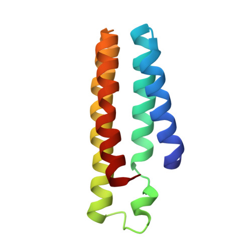
    Proteins represent the most sophisticated building blocks available to an organism and to the laboratory chemist. Yet, in contrast to nearly all other types of molecular building blocks, the designed self-assembly of proteins has largely been inaccessible because of the chemical and structural heterogeneity of protein surfaces. To circumvent the challenge of programming extensive non-covalent interactions to control protein self-assembly, we have previously exploited the directionality and strength of metal coordination interactions to guide the formation of closed, homoligomeric protein assemblies. Here, we extend this strategy to the generation of periodic protein arrays. We show that a monomeric protein with properly oriented coordination motifs on its surface can arrange, on metal binding, into one-dimensional nanotubes and two- or three-dimensional crystalline arrays with dimensions that collectively span nearly the entire nano- and micrometre scale. The assembly of these arrays is tuned predictably by external stimuli, such as metal concentration and pH.


  • Organizational Affiliation

    Department of Chemistry and Biochemistry, University of California, San Diego, La Jolla, California 92093, USA.


Macromolecules
Find similar proteins by:  (by identity cutoff)  |  3D Structure
Entity ID: 1
MoleculeChains Sequence LengthOrganismDetailsImage
Soluble cytochrome b562
A, B, C, D
106Escherichia coliMutation(s): 14 
Gene Names: cybC
UniProt
Find proteins for P0ABE7 (Escherichia coli)
Explore P0ABE7 
Go to UniProtKB:  P0ABE7
Entity Groups  
Sequence Clusters30% Identity50% Identity70% Identity90% Identity95% Identity100% Identity
UniProt GroupP0ABE7
Sequence Annotations
Expand
  • Reference Sequence
Small Molecules
Ligands 2 Unique
IDChains Name / Formula / InChI Key2D Diagram3D Interactions
HEM
Query on HEM

Download Ideal Coordinates CCD File 
E [auth A],
G [auth B],
J [auth C],
L [auth D]
PROTOPORPHYRIN IX CONTAINING FE
C34 H32 Fe N4 O4
KABFMIBPWCXCRK-RGGAHWMASA-L
ZN
Query on ZN

Download Ideal Coordinates CCD File 
F [auth A]
H [auth B]
I [auth B]
K [auth C]
M [auth D]
F [auth A],
H [auth B],
I [auth B],
K [auth C],
M [auth D],
N [auth D]
ZINC ION
Zn
PTFCDOFLOPIGGS-UHFFFAOYSA-N
Experimental Data & Validation

Experimental Data

  • Method: X-RAY DIFFRACTION
  • Resolution: 2.30 Å
  • R-Value Free: 0.286 
  • R-Value Work: 0.210 
  • R-Value Observed: 0.215 
  • Space Group: C 1 2 1
Unit Cell:
Length ( Å )Angle ( ˚ )
a = 95.69α = 90
b = 37.843β = 112.61
c = 138.489γ = 90
Software Package:
Software NamePurpose
MOSFLMdata reduction
SCALAdata scaling
PHASERphasing
REFMACrefinement
PDB_EXTRACTdata extraction
Blu-Icedata collection

Structure Validation

View Full Validation Report


Ligand Structure Quality Assessment 


Entry History 

Deposition Data

Revision History  (Full details and data files)

  • Version 1.0: 2012-07-04
    Type: Initial release
  • Version 1.1: 2024-02-28
    Changes: Data collection, Database references, Derived calculations, Refinement description