3SZR

Crystal structure of modified nucleotide-free human MxA


Experimental Data Snapshot

  • Method: X-RAY DIFFRACTION
  • Resolution: 3.50 Å
  • R-Value Free: 0.295 
  • R-Value Work: 0.262 
  • R-Value Observed: 0.263 

wwPDB Validation   3D Report Full Report


This is version 1.4 of the entry. See complete history


Literature

Structure of myxovirus resistance protein a reveals intra- and intermolecular domain interactions required for the antiviral function.

Gao, S.von der Malsburg, A.Dick, A.Faelber, K.Schroder, G.F.Haller, O.Kochs, G.Daumke, O.

(2011) Immunity 35: 514-525

  • DOI: https://doi.org/10.1016/j.immuni.2011.07.012
  • Primary Citation of Related Structures:  
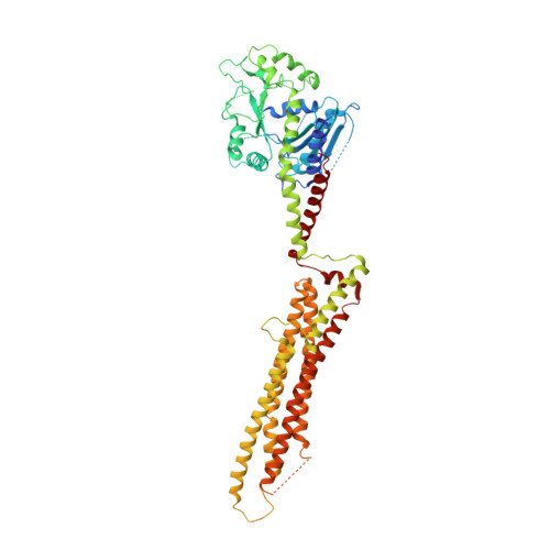
    3SZR

  • PubMed Abstract: 

    Human myxovirus resistance protein 1 (MxA) is an interferon-induced dynamin-like GTPase that acts as a cell-autonomous host restriction factor against many viral pathogens including influenza viruses. To study the molecular principles of its antiviral activity, we determined the crystal structure of nucleotide-free MxA, which showed an extended three-domain architecture. The central bundle signaling element (BSE) connected the amino-terminal GTPase domain with the stalk via two hinge regions. MxA oligomerized in the crystal via the stalk and the BSE, which in turn interacted with the stalk of the neighboring monomer. We demonstrated that the intra- and intermolecular domain interplay between the BSE and stalk was essential for oligomerization and the antiviral function of MxA. Based on these results, we propose a structural model for the mechano-chemical coupling in ring-like MxA oligomers as the principle mechanism for this unique antiviral effector protein.


  • Organizational Affiliation

    Max-Delbrück-Centrum for Molecular Medicine, Crystallography, Robert-Rössle-Strasse 10, 13125 Berlin, Germany. song.gao@mdc-berlin.de


Macromolecules
Find similar proteins by:  (by identity cutoff)  |  3D Structure
Entity ID: 1
MoleculeChains Sequence LengthOrganismDetailsImage
Interferon-induced GTP-binding protein Mx1608Homo sapiensMutation(s): 4 
Gene Names: MX1
UniProt & NIH Common Fund Data Resources
Find proteins for P20591 (Homo sapiens)
Explore P20591 
Go to UniProtKB:  P20591
PHAROS:  P20591
GTEx:  ENSG00000157601 
Entity Groups  
Sequence Clusters30% Identity50% Identity70% Identity90% Identity95% Identity100% Identity
UniProt GroupP20591
Sequence Annotations
Expand
  • Reference Sequence
Experimental Data & Validation

Experimental Data

  • Method: X-RAY DIFFRACTION
  • Resolution: 3.50 Å
  • R-Value Free: 0.295 
  • R-Value Work: 0.262 
  • R-Value Observed: 0.263 
  • Space Group: C 1 2 1
Unit Cell:
Length ( Å )Angle ( ˚ )
a = 156.82α = 90
b = 134.08β = 106.34
c = 58.1γ = 90
Software Package:
Software NamePurpose
REFMACrefinement
PHASERphasing
CNSrefinement
iMOSFLMdata reduction
XDSdata reduction
XDSdata scaling

Structure Validation

View Full Validation Report


Entry History 

Deposition Data

Revision History  (Full details and data files)

  • Version 1.0: 2011-10-12
    Type: Initial release
  • Version 1.1: 2011-10-26
    Changes: Database references
  • Version 1.2: 2011-11-16
    Changes: Database references
  • Version 1.3: 2019-07-17
    Changes: Data collection, Refinement description
  • Version 1.4: 2023-09-13
    Changes: Data collection, Database references, Refinement description