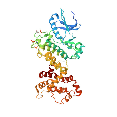
Cofactor-mediated conformational control in the bifunctional kinase/RNase Ire1.
Korennykh, A.V., Egea, P.F., Korostelev, A.A., Finer-Moore, J., Stroud, R.M., Zhang, C., Shokat, K.M., Walter, P.(2011) BMC Biol 9: 48-48
- PubMed: 21729334
- DOI: https://doi.org/10.1186/1741-7007-9-48
- Primary Citation of Related Structures:
3SDM - PubMed Abstract:
Ire1 is a signal transduction protein in the endoplasmic reticulum (ER) membrane that serves to adjust the protein-folding capacity of the ER according to the needs of the cell. Ire1 signals, in a transcriptional program, the unfolded protein response (UPR) via the coordinated action of its protein kinase and RNase domains. In this study, we investigated how the binding of cofactors to the kinase domain of Ire1 modulates its RNase activity. Our results suggest that the kinase domain of Ire1 initially binds cofactors without activation of the RNase domain. RNase is activated upon a subsequent conformational rearrangement of Ire1 governed by the chemical properties of bound cofactors. The conformational step can be selectively inhibited by chemical perturbations of cofactors. Substitution of a single oxygen atom in the terminal β-phosphate group of a potent cofactor ADP by sulfur results in ADPβS, a cofactor that binds to Ire1 as well as to ADP but does not activate RNase. RNase activity can be rescued by thiophilic metal ions such as Mn2+ and Cd2+, revealing a functional metal ion-phosphate interaction which controls the conformation and RNase activity of the Ire1 ADP complex. Mutagenesis of the kinase domain suggests that this rearrangement involves movement of the αC-helix, which is generally conserved among protein kinases. Using X-ray crystallography, we show that oligomerization of Ire1 is sufficient for placing the αC-helix in the active, cofactor-bound-like conformation, even in the absence of cofactors. Our structural and biochemical evidence converges on a model that the cofactor-induced conformational change in Ire1 is coupled to oligomerization of the receptor, which, in turn, activates RNase. The data reveal that cofactor-Ire1 interactions occur in two independent steps: binding of a cofactor to Ire1 and subsequent rearrangement of Ire1 resulting in its self-association. The pronounced allosteric effect of cofactors on protein-protein interactions involving Ire1's kinase domain suggests that protein kinases and pseudokinases encoded in metazoan genomes may use ATP pocket-binding ligands similarly to exert signaling roles other than phosphoryl transfer.
Organizational Affiliation:
Howard Hughes Medical Institute, University of California, San Francisco, 600 16th Street, Room S272, Box 0724, San Francisco, CA 94158, USA. akorenny@princeton.edu