3SC6

2.65 Angstrom resolution crystal structure of dTDP-4-dehydrorhamnose reductase (rfbD) from Bacillus anthracis str. Ames in complex with NADP


Experimental Data Snapshot

  • Method: X-RAY DIFFRACTION
  • Resolution: 2.65 Å
  • R-Value Free: 0.258 
  • R-Value Work: 0.215 
  • R-Value Observed: 0.217 

wwPDB Validation   3D Report Full Report


Ligand Structure Quality Assessment 


This is version 1.5 of the entry. See complete history


Literature

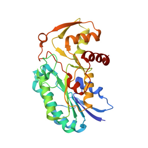
Structure of the Bacillus anthracis dTDP-L-rhamnose-biosynthetic enzyme dTDP-4-dehydrorhamnose reductase (RfbD).

Law, A.Stergioulis, A.Halavaty, A.S.Minasov, G.Anderson, W.F.Kuhn, M.L.

(2017) Acta Crystallogr F Struct Biol Commun 73: 644-650

  • DOI: https://doi.org/10.1107/S2053230X17015746
  • Primary Citation of Related Structures:  
    3SC6

  • PubMed Abstract: 

    Bacillus anthracis is the causative agent of the deadly disease Anthrax. Its use in bioterrorism and its ability to re-emerge have brought renewed interest in this organism. B. anthracis is a Gram-positive bacterium that adds L-rhamnose to its cell-wall polysaccharides using the activated donor dTDP-β-L-rhamnose. The enzymes involved in the biosynthesis of the activated donor are absent in humans, which make them ideal targets for therapeutic development to combat pathogens. Here, the 2.65 Å resolution crystal structure of the fourth enzyme in the dTDP-β-L-rhamnose-biosynthetic pathway from B. anthracis, dTDP-4-dehydro-β-L-rhamnose reductase (RfbD), is presented in complex with NADP + . This enzyme catalyzes the reduction of dTDP-4-dehydro-β-L-rhamnose to dTDP-β-L-rhamnose. Although the protein was co-crystallized in the presence of Mg 2+ , the protein lacks the conserved residues that coordinate Mg 2+ .


  • Organizational Affiliation

    Department of Chemistry and Biochemistry, San Francisco State University, USA.


Macromolecules
Find similar proteins by:  (by identity cutoff)  |  3D Structure
Entity ID: 1
MoleculeChains Sequence LengthOrganismDetailsImage
dTDP-4-dehydrorhamnose reductase
A, B, C, D, E
A, B, C, D, E, F
287Bacillus anthracis str. AmesMutation(s): 0 
Gene Names: BAS1138BA_1231GBAA_1231rfbD
EC: 1.1.1.133
UniProt
Find proteins for A0A6L7H7R4 (Bacillus anthracis)
Explore A0A6L7H7R4 
Go to UniProtKB:  A0A6L7H7R4
Entity Groups  
Sequence Clusters30% Identity50% Identity70% Identity90% Identity95% Identity100% Identity
UniProt GroupA0A6L7H7R4
Sequence Annotations
Expand
  • Reference Sequence
Small Molecules
Ligands 2 Unique
IDChains Name / Formula / InChI Key2D Diagram3D Interactions
NAP
Query on NAP

Download Ideal Coordinates CCD File 
G [auth A]
J [auth B]
O [auth C]
R [auth D]
U [auth E]
G [auth A],
J [auth B],
O [auth C],
R [auth D],
U [auth E],
Z [auth F]
NADP NICOTINAMIDE-ADENINE-DINUCLEOTIDE PHOSPHATE
C21 H28 N7 O17 P3
XJLXINKUBYWONI-NNYOXOHSSA-N
SO4
Query on SO4

Download Ideal Coordinates CCD File 
AA [auth F]
BA [auth F]
CA [auth F]
H [auth A]
I [auth A]
AA [auth F],
BA [auth F],
CA [auth F],
H [auth A],
I [auth A],
K [auth B],
L [auth B],
M [auth B],
N [auth B],
P [auth C],
Q [auth C],
S [auth D],
T [auth D],
V [auth E],
W [auth E],
X [auth E],
Y [auth E]
SULFATE ION
O4 S
QAOWNCQODCNURD-UHFFFAOYSA-L
Experimental Data & Validation

Experimental Data

Unit Cell:
Length ( Å )Angle ( ˚ )
a = 79.449α = 90
b = 113.337β = 91.18
c = 144.949γ = 90
Software Package:
Software NamePurpose
Blu-Icedata collection
PHASERphasing
REFMACrefinement
HKL-2000data reduction
HKL-2000data scaling

Structure Validation

View Full Validation Report


Ligand Structure Quality Assessment 


Entry History 

Revision History  (Full details and data files)

  • Version 1.0: 2011-06-22
    Type: Initial release
  • Version 1.1: 2011-07-13
    Changes: Version format compliance
  • Version 1.2: 2017-08-23
    Changes: Experimental preparation
  • Version 1.3: 2017-11-08
    Changes: Refinement description
  • Version 1.4: 2017-12-13
    Changes: Database references
  • Version 1.5: 2023-09-13
    Changes: Data collection, Database references, Derived calculations, Refinement description