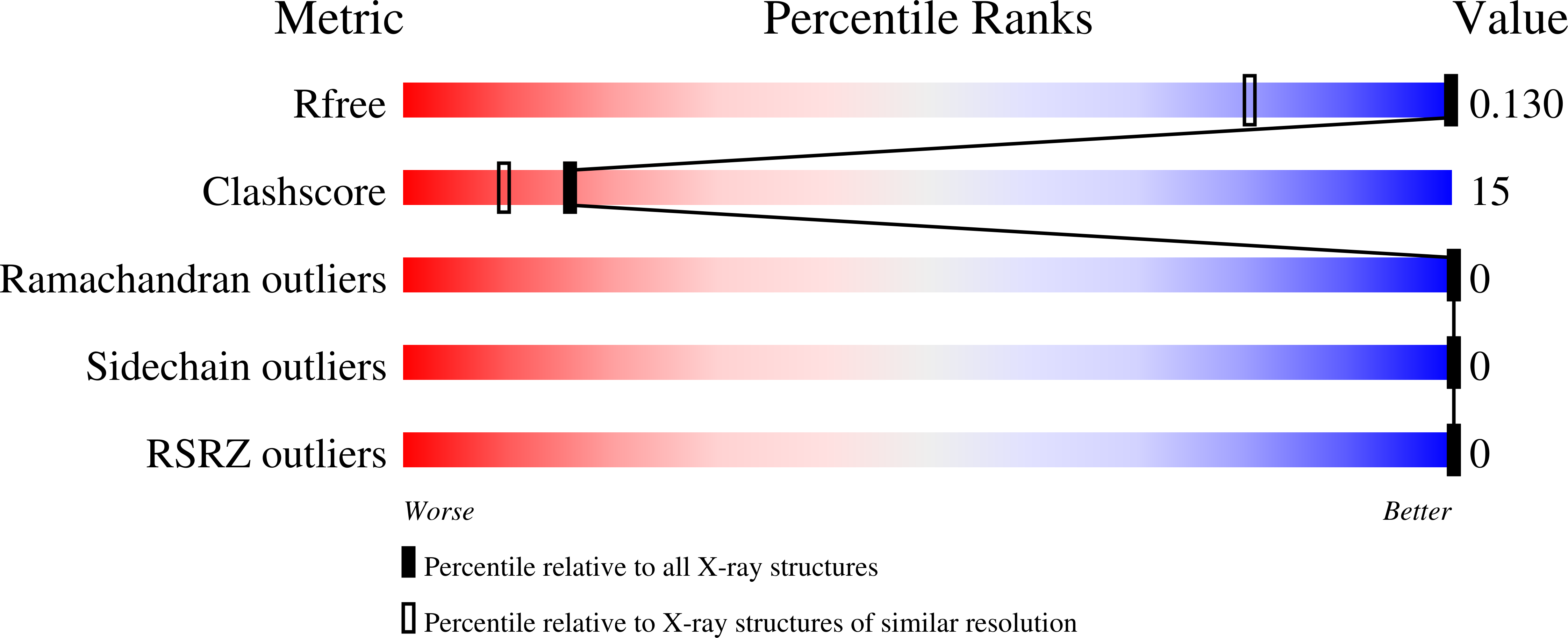
3SBN

trichovirin I-4A in polar environment at 0.9 Angstroem


Experimental Data Snapshot

  • Method: X-RAY DIFFRACTION
  • Resolution: 0.90 Å
  • R-Value Free: 0.128 
  • R-Value Observed: 0.102 

wwPDB Validation   3D Report Full Report


This is version 2.0 of the entry. See complete history


Literature

Four complete turns of a curved 310-helix at atomic resolution: The crystal structure of the peptaibol trichovirin I-4A in polar environment suggests a transition to alpha-helix for membrane function

Gessmann, R.Axford, D.Owen, R.L.Bruckner, H.Petratos, K.

(2012) Acta Crystallogr D Biol Crystallogr 68: 109-116

  • DOI: https://doi.org/10.1107/S090744491105133X
  • Primary Citation of Related Structures:  
    3SBN

  • PubMed Abstract: 

    The first crystal structure of a member of peptaibol antibiotic subfamily 4, trichovirin I-4A (14 residues), has been determined by direct methods and refined at atomic resolution. The monoclinic unit cell has two molecules in the asymmetric unit. Both molecules assume a 3₁₀ right-handed helical conformation and are significantly bent. The molecules pack loosely along the crystallographic twofold axis, forming two large tunnels between symmetry-related molecules in which no ordered solvent could be located. Carbonyl O atoms which are not involved in intramolecular hydrogen bonding participate in close van der Waals interactions with apolar groups. The necessary amphipathicity for biological activity of peptaibols is not realised in the crystal structure. Hence, a structural change of trichovirin to an α-helical conformation is proposed for membrane integration and efficient water/ion transportation across the lipid bilayer.


  • Organizational Affiliation

    IMBB-FORTH, N. Plastira 100, Heraklion, Crete 70013, Greece.


Macromolecules

Find similar proteins by:  Sequence   |   3D Structure  

Entity ID: 1
MoleculeChains Sequence LengthOrganismDetailsImage
Trichovirin I-4A
A, B
15Trichoderma virideMutation(s): 0 
Entity Groups  
Sequence Clusters30% Identity50% Identity70% Identity90% Identity95% Identity100% Identity
Sequence Annotations
Expand
  • Reference Sequence
Biologically Interesting Molecules (External Reference) 1 Unique
Experimental Data & Validation

Experimental Data

  • Method: X-RAY DIFFRACTION
  • Resolution: 0.90 Å
  • R-Value Free: 0.128 
  • R-Value Observed: 0.102 
  • Space Group: P 1 21 1
Unit Cell:
Length ( Å )Angle ( ˚ )
a = 24.284α = 90
b = 9.896β = 97.15
c = 37.567γ = 90
Software Package:
Software NamePurpose
GDAdata collection
ACORNphasing
SHELXLrefinement
XDSdata reduction
SCALAdata scaling

Structure Validation

View Full Validation Report


Entry History 

Deposition Data

Revision History  (Full details and data files)

  • Version 1.0: 2011-12-28
    Type: Initial release
  • Version 1.1: 2012-02-15
    Changes: Database references
  • Version 1.2: 2012-12-12
    Changes: Other
  • Version 2.0: 2023-11-15
    Changes: Atomic model, Data collection, Database references, Derived calculations