3S9E

Crystal structure of the kainate receptor GluK3 ligand binding domain in complex with (S)-glutamate


Experimental Data Snapshot

  • Method: X-RAY DIFFRACTION
  • Resolution: 1.60 Å
  • R-Value Free: 0.184 
  • R-Value Work: 0.135 
  • R-Value Observed: 0.138 

wwPDB Validation   3D Report Full Report


This is version 1.5 of the entry. See complete history


Literature

Binding site and interlobe interactions of the ionotropic glutamate receptor GluK3 ligand binding domain revealed by high resolution crystal structure in complex with (S)-glutamate.

Venskutonyte, R.Frydenvang, K.Gajhede, M.Bunch, L.Pickering, D.S.Kastrup, J.S.

(2011) J Struct Biol 176: 307-314

  • DOI: https://doi.org/10.1016/j.jsb.2011.08.014
  • Primary Citation of Related Structures:  
    3S9E, 4MH5

  • PubMed Abstract: 

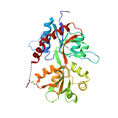
    Ionotropic glutamate receptors (iGluRs) are involved in excitatory signal transmission throughout the central nervous system and their malfunction is associated with various health disorders. GluK3 is a subunit of iGluRs, belonging to the subfamily of kainate receptors (GluK1-5). Several crystal structures of GluK1 and GluK2 ligand binding domains have been determined in complex with agonists and antagonists. However, little is known about the molecular mechanisms underlying GluK3 ligand binding properties and no compounds displaying reasonable selectivity towards GluK3 are available today. Here, we present the first X-ray crystal structure of the ligand binding domain of GluK3 in complex with glutamate, determined to 1.6Å resolution. The structure reveals a conserved glutamate binding mode, characteristic for iGluRs, and a water molecule network in the glutamate binding site similar to that seen in GluK1. In GluK3, a slightly lower degree of domain closure around glutamate is observed compared to most other kainate receptor structures with glutamate. The volume of the GluK3 glutamate binding cavity was found to be of intermediate size between those of GluK1 and GluK2. The residues in GluK3 contributing to the subfamily differences in the binding sites are primarily: Thr520, Ala691, Asn722, Leu736 and Thr742. The GluK3 ligand binding domain seems to be less stabilized through interlobe interactions than GluK1 and this may contribute to the faster desensitization kinetics of GluK3.


  • Organizational Affiliation

    Department of Medicinal Chemistry, University of Copenhagen, Universitetsparken 2, DK-2100 Copenhagen, Denmark.


Macromolecules
Find similar proteins by:  (by identity cutoff)  |  3D Structure
Entity ID: 1
MoleculeChains Sequence LengthOrganismDetailsImage
Glutamate receptor, ionotropic kainate 3
A, B
258Rattus norvegicusMutation(s): 0 
Gene Names: Grik3
UniProt
Find proteins for P42264 (Rattus norvegicus)
Explore P42264 
Go to UniProtKB:  P42264
Entity Groups  
Sequence Clusters30% Identity50% Identity70% Identity90% Identity95% Identity100% Identity
UniProt GroupP42264
Sequence Annotations
Expand
  • Reference Sequence
Small Molecules
Ligands 5 Unique
IDChains Name / Formula / InChI Key2D Diagram3D Interactions
GLU
Query on GLU

Download Ideal Coordinates CCD File 
C [auth A],
J [auth B]
GLUTAMIC ACID
C5 H9 N O4
WHUUTDBJXJRKMK-VKHMYHEASA-N
PO4
Query on PO4

Download Ideal Coordinates CCD File 
G [auth A],
H [auth A],
M [auth B]
PHOSPHATE ION
O4 P
NBIIXXVUZAFLBC-UHFFFAOYSA-K
GOL
Query on GOL

Download Ideal Coordinates CCD File 
D [auth A]GLYCEROL
C3 H8 O3
PEDCQBHIVMGVHV-UHFFFAOYSA-N
CL
Query on CL

Download Ideal Coordinates CCD File 
E [auth A],
F [auth A],
K [auth B],
L [auth B]
CHLORIDE ION
Cl
VEXZGXHMUGYJMC-UHFFFAOYSA-M
NA
Query on NA

Download Ideal Coordinates CCD File 
I [auth A],
N [auth B],
O [auth B]
SODIUM ION
Na
FKNQFGJONOIPTF-UHFFFAOYSA-N
Binding Affinity Annotations 
IDSourceBinding Affinity
GLU PDBBind:  3S9E Ki: 7430 (nM) from 1 assay(s)
Experimental Data & Validation

Experimental Data

  • Method: X-RAY DIFFRACTION
  • Resolution: 1.60 Å
  • R-Value Free: 0.184 
  • R-Value Work: 0.135 
  • R-Value Observed: 0.138 
  • Space Group: P 41
Unit Cell:
Length ( Å )Angle ( ˚ )
a = 67.8α = 90
b = 67.8β = 90
c = 122.43γ = 90
Software Package:
Software NamePurpose
MAR345data collection
PHASERphasing
PHENIXrefinement
MOSFLMdata reduction
SCALAdata scaling

Structure Validation

View Full Validation Report


Entry History 

Deposition Data

Revision History  (Full details and data files)

  • Version 1.0: 2011-09-28
    Type: Initial release
  • Version 1.1: 2012-05-23
    Changes: Database references
  • Version 1.2: 2013-10-02
    Changes: Other
  • Version 1.3: 2017-08-09
    Changes: Refinement description, Source and taxonomy
  • Version 1.4: 2017-11-08
    Changes: Refinement description
  • Version 1.5: 2023-09-13
    Changes: Data collection, Database references, Derived calculations, Refinement description