3RZZ

Structure of Hydroxyethylphoshphonate Dioxygenase Y98F Mutant


Experimental Data Snapshot

  • Method: X-RAY DIFFRACTION
  • Resolution: 2.20 Å
  • R-Value Free: 0.268 
  • R-Value Work: 0.207 

wwPDB Validation   3D Report Full Report


This is version 1.2 of the entry. See complete history


Literature

Mechanism and substrate recognition of 2-hydroxyethylphosphonate dioxygenase.

Peck, S.C.Cooke, H.A.Cicchillo, R.M.Malova, P.Hammerschmidt, F.Nair, S.K.van der Donk, W.A.

(2011) Biochemistry 50: 6598-6605

  • DOI: https://doi.org/10.1021/bi200804r
  • Primary Citation of Related Structures:  
    3RZZ

  • PubMed Abstract: 

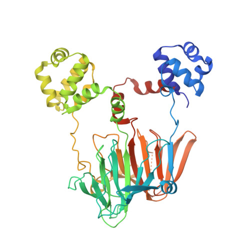
    HEPD belongs to the superfamily of 2-His-1-carboxylate non-heme iron-dependent dioxygenases. It converts 2-hydroxyethylphosphonate (2-HEP) to hydroxymethylphosphonate (HMP) and formate. Previously postulated mechanisms for the reaction catalyzed by HEPD cannot explain its conversion of 1-HEP to acetylphosphate. Alternative mechanisms that involve either phosphite or methylphosphonate as intermediates, which potentially explain all experimental studies including isotope labeling experiments and use of substrate analogues, were investigated. The results of these studies reveal that these alternative mechanisms are not correct. Site-directed mutagenesis studies of Lys16, Arg90, and Tyr98 support roles of these residues in binding of 2-HEP. Mutation of Lys16 to Ala resulted in an inactive enzyme, whereas mutation of Arg90 to Ala or Tyr98 to Phe greatly decreased k(cat)/K(m,2-HEP). Furthermore, the latter mutants could not be saturated in O(2). These results suggest that proper binding of 2-HEP is important for O(2) activation and that the enzyme uses a compulsory binding order with 2-HEP binding before O(2). The Y98F mutant produces methylphosphonate as a minor side product providing indirect support for the proposal that the last step during catalysis involves a ferric hydroxide reacting with a methylphosphonate radical.


  • Organizational Affiliation

    Department of Chemistry and Howard Hughes Medical Institute and ‡Institute for Genomic Biology, University of Illinois , 1206 West Gregory Drive, Urbana, Illinois 61801, United States.


Macromolecules
Find similar proteins by:  (by identity cutoff)  |  3D Structure
Entity ID: 1
MoleculeChains Sequence LengthOrganismDetailsImage
Hydroxyethylphoshphonate Dioxygenase (PhpD)446Streptomyces viridochromogenesMutation(s): 1 
Gene Names: phpD
UniProt
Find proteins for Q5IW40 (Streptomyces viridochromogenes (strain DSM 40736 / JCM 4977 / BCRC 1201 / Tue 494))
Explore Q5IW40 
Go to UniProtKB:  Q5IW40
Entity Groups  
Sequence Clusters30% Identity50% Identity70% Identity90% Identity95% Identity100% Identity
UniProt GroupQ5IW40
Sequence Annotations
Expand
  • Reference Sequence
Small Molecules
Ligands 1 Unique
IDChains Name / Formula / InChI Key2D Diagram3D Interactions
CD
Query on CD

Download Ideal Coordinates CCD File 
B [auth A]
C [auth A]
D [auth A]
E [auth A]
F [auth A]
B [auth A],
C [auth A],
D [auth A],
E [auth A],
F [auth A],
G [auth A],
H [auth A],
I [auth A],
J [auth A],
K [auth A],
L [auth A],
M [auth A],
N [auth A],
O [auth A],
P [auth A]
CADMIUM ION
Cd
WLZRMCYVCSSEQC-UHFFFAOYSA-N
Experimental Data & Validation

Experimental Data

  • Method: X-RAY DIFFRACTION
  • Resolution: 2.20 Å
  • R-Value Free: 0.268 
  • R-Value Work: 0.207 
  • Space Group: C 2 2 21
Unit Cell:
Length ( Å )Angle ( ˚ )
a = 84.747α = 90
b = 94.964β = 90
c = 101.396γ = 90
Software Package:
Software NamePurpose
HKL-2000data collection
PHASERphasing
CNSrefinement
HKL-2000data reduction
HKL-2000data scaling

Structure Validation

View Full Validation Report


Entry History 

Deposition Data

  • Released Date: 2011-07-20 
  • Deposition Author(s): Cooke, H.A.

Revision History  (Full details and data files)

  • Version 1.0: 2011-07-20
    Type: Initial release
  • Version 1.1: 2012-05-23
    Changes: Database references
  • Version 1.2: 2023-09-13
    Changes: Data collection, Database references, Derived calculations, Refinement description