3RIL

The acid beta-glucosidase active site exhibits plasticity in binding 3,4,5,6-tetrahydroxyazepane-based inhibitors: implications for pharmacological chaperone design for gaucher disease


Experimental Data Snapshot

  • Method: X-RAY DIFFRACTION
  • Resolution: 2.40 Å
  • R-Value Free: 0.232 
  • R-Value Work: 0.171 
  • R-Value Observed: 0.174 

wwPDB Validation   3D Report Full Report


Ligand Structure Quality Assessment 


This is version 1.3 of the entry. See complete history


Literature

Binding of 3,4,5,6-tetrahydroxyazepanes to the acid-beta-glucosidase active site: implications for pharmacological chaperone design for Gaucher disease

Orwig, S.D.Tan, Y.L.Grimster, N.P.Yu, Z.Powers, E.T.Kelly, J.W.Lieberman, R.L.

(2011) Biochemistry 50: 10647-10657

  • DOI: https://doi.org/10.1021/bi201619z
  • Primary Citation of Related Structures:  
    3RIK, 3RIL

  • PubMed Abstract: 

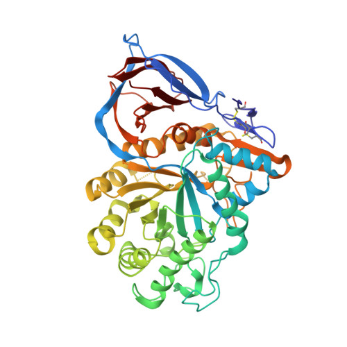
    Pharmacologic chaperoning is a therapeutic strategy being developed to improve the cellular folding and trafficking defects associated with Gaucher disease, a lysosomal storage disorder caused by point mutations in the gene encoding acid-β-glucosidase (GCase). In this approach, small molecules bind to and stabilize mutant folded or nearly folded GCase in the endoplasmic reticulum (ER), increasing the concentration of folded, functional GCase trafficked to the lysosome where the mutant enzyme can hydrolyze the accumulated substrate. To date, the pharmacologic chaperone (PC) candidates that have been investigated largely have been active site-directed inhibitors of GCase, usually containing five- or six-membered rings, such as modified azasugars. Here we show that a seven-membered, nitrogen-containing heterocycle (3,4,5,6-tetrahydroxyazepane) scaffold is also promising for generating PCs for GCase. Crystal structures reveal that the core azepane stabilizes GCase in a variation of its proposed active conformation, whereas binding of an analogue with an N-linked hydroxyethyl tail stabilizes GCase in a conformation in which the active site is covered, also utilizing a loop conformation not seen previously. Although both compounds preferentially stabilize GCase to thermal denaturation at pH 7.4, reflective of the pH in the ER, only the core azepane, which is a mid-micromolar competitive inhibitor, elicits a modest increase in enzyme activity for the neuronopathic G202R and the non-neuronopathic N370S mutant GCase in an intact cell assay. Our results emphasize the importance of the conformational variability of the GCase active site in the design of competitive inhibitors as PCs for Gaucher disease.


  • Organizational Affiliation

    School of Chemistry and Biochemistry, Georgia Institute of Technology, Atlanta, Georgia 30332, United States.


Macromolecules
Find similar proteins by:  (by identity cutoff)  |  3D Structure
Entity ID: 1
MoleculeChains Sequence LengthOrganismDetailsImage
Glucosylceramidase
A, B, C, D
497Homo sapiensMutation(s): 1 
Gene Names: GBAGCGLUC
EC: 3.2.1.45
UniProt & NIH Common Fund Data Resources
Find proteins for P04062 (Homo sapiens)
Explore P04062 
Go to UniProtKB:  P04062
PHAROS:  P04062
GTEx:  ENSG00000177628 
Entity Groups  
Sequence Clusters30% Identity50% Identity70% Identity90% Identity95% Identity100% Identity
UniProt GroupP04062
Sequence Annotations
Expand
  • Reference Sequence
Small Molecules
Ligands 3 Unique
IDChains Name / Formula / InChI Key2D Diagram3D Interactions
NAG
Query on NAG

Download Ideal Coordinates CCD File 
LA [auth C],
P [auth A],
UA [auth D],
Y [auth B]
2-acetamido-2-deoxy-beta-D-glucopyranose
C8 H15 N O6
OVRNDRQMDRJTHS-FMDGEEDCSA-N
3RK
Query on 3RK

Download Ideal Coordinates CCD File 
MA [auth C],
Q [auth A],
VA [auth D],
Z [auth B]
(3S,4R,5R,6S)-azepane-3,4,5,6-tetrol
C6 H13 N O4
MRFFNLOQLBWKPJ-UNTFVMJOSA-N
SO4
Query on SO4

Download Ideal Coordinates CCD File 
AA [auth C]
BA [auth C]
CA [auth C]
DA [auth C]
E [auth A]
AA [auth C],
BA [auth C],
CA [auth C],
DA [auth C],
E [auth A],
EA [auth C],
F [auth A],
FA [auth C],
G [auth A],
GA [auth C],
H [auth A],
HA [auth C],
I [auth A],
IA [auth C],
J [auth A],
JA [auth C],
K [auth A],
KA [auth C],
L [auth A],
M [auth A],
N [auth A],
NA [auth D],
O [auth A],
OA [auth D],
PA [auth D],
QA [auth D],
R [auth B],
RA [auth D],
S [auth B],
SA [auth D],
T [auth B],
TA [auth D],
U [auth B],
V [auth B],
W [auth B],
X [auth B]
SULFATE ION
O4 S
QAOWNCQODCNURD-UHFFFAOYSA-L
Binding Affinity Annotations 
IDSourceBinding Affinity
3RK BindingDB:  3RIL Ki: 1.28e+4 (nM) from 1 assay(s)
PDBBind:  3RIL IC50: 1.46e+5 (nM) from 1 assay(s)
Binding MOAD:  3RIL IC50: 1.46e+5 (nM) from 1 assay(s)
Experimental Data & Validation

Experimental Data

  • Method: X-RAY DIFFRACTION
  • Resolution: 2.40 Å
  • R-Value Free: 0.232 
  • R-Value Work: 0.171 
  • R-Value Observed: 0.174 
  • Space Group: P 1 21 1
Unit Cell:
Length ( Å )Angle ( ˚ )
a = 109.217α = 90
b = 91.425β = 110.95
c = 152.659γ = 90
Software Package:
Software NamePurpose
SERGUIdata collection
REFMACrefinement
HKL-2000data reduction
HKL-2000data scaling
REFMACphasing

Structure Validation

View Full Validation Report


Ligand Structure Quality Assessment 


Entry History 

Deposition Data

Revision History  (Full details and data files)

  • Version 1.0: 2012-03-14
    Type: Initial release
  • Version 1.1: 2012-12-05
    Changes: Derived calculations
  • Version 1.2: 2018-08-15
    Changes: Advisory, Data collection, Derived calculations
  • Version 1.3: 2020-07-29
    Type: Remediation
    Reason: Carbohydrate remediation
    Changes: Data collection, Database references, Derived calculations, Structure summary