


Crystal structure of a Hypothetical sugar kinase (CHU_1875) from CYTOPHAGA HUTCHINSONII ATCC 33406 at 1.65 A resolution
Joint Center for Structural Genomics (JCSG)To be published.
Experimental Data Snapshot
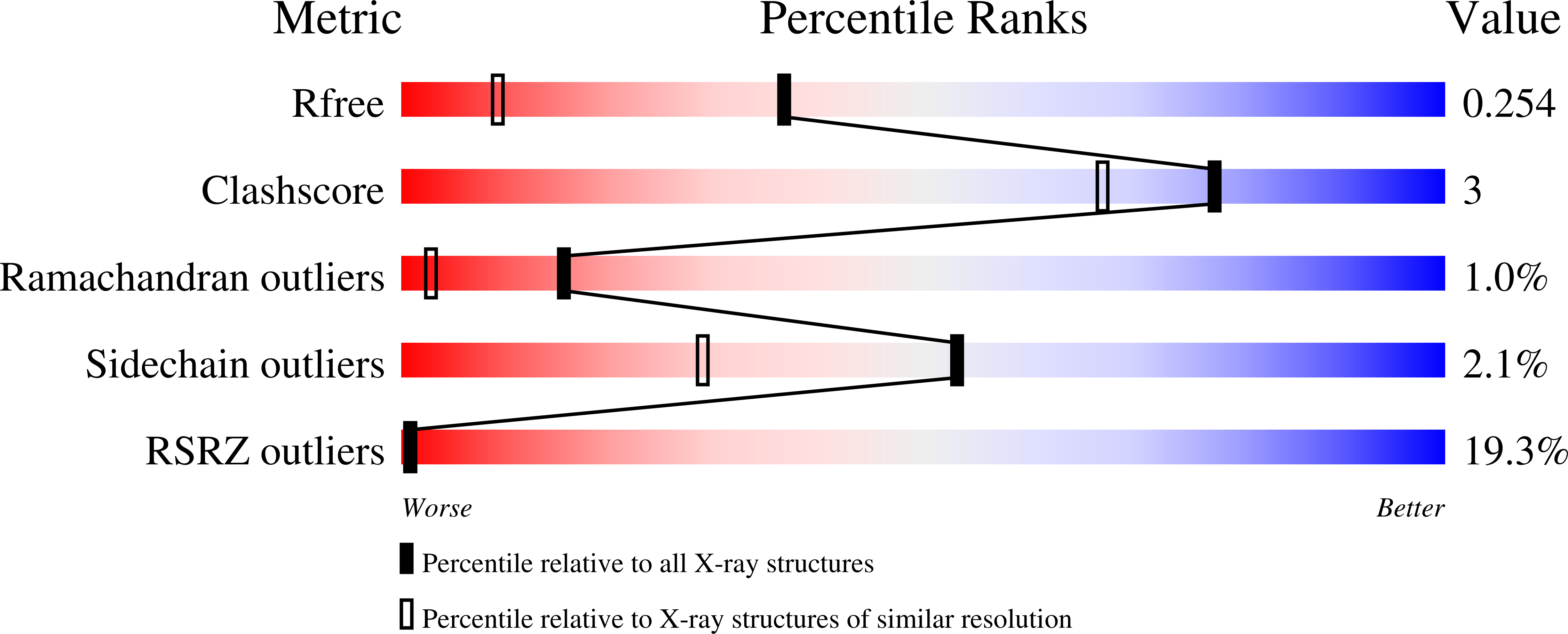
wwPDB Validation 3D Report Full Report
Entity ID: 1 | |||||
|---|---|---|---|---|---|
| Molecule | Chains | Sequence Length | Organism | Details | Image |
| Hypothetical sugar kinase | 321 | Cytophaga hutchinsonii ATCC 33406 | Mutation(s): 0 Gene Names: glk, CHU_1875 | 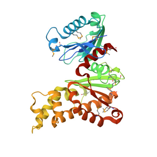 | |
Entity Groups | |||||
| Sequence Clusters | 30% Identity50% Identity70% Identity90% Identity95% Identity100% Identity | ||||
Sequence AnnotationsExpand | |||||
| |||||
| Modified Residues 1 Unique | |||||
|---|---|---|---|---|---|
| ID | Chains | Type | Formula | 2D Diagram | Parent |
| MSE Query on MSE | A | L-PEPTIDE LINKING | C5 H11 N O2 Se |  | MET |
| Length ( Å ) | Angle ( ˚ ) |
|---|---|
| a = 71.827 | α = 90 |
| b = 78.359 | β = 90 |
| c = 65.744 | γ = 90 |
| Software Name | Purpose |
|---|---|
| MolProbity | model building |
| PDB_EXTRACT | data extraction |
| SOLVE | phasing |
| SCALA | data scaling |
| BUSTER-TNT | refinement |
| Xpleo | model building |
| MOSFLM | data reduction |
| BUSTER | refinement |
RCSB PDB (citation) is hosted by
RCSB PDB is a member of the

