3QXR

Crystal structure of the brominated CKIT-1 proto-oncogene promoter quadruplex DNA


Experimental Data Snapshot

  • Method: X-RAY DIFFRACTION
  • Resolution: 1.62 Å
  • R-Value Free: 0.213 
  • R-Value Work: 0.166 
  • R-Value Observed: 0.168 

wwPDB Validation   3D Report Full Report


This is version 1.5 of the entry. See complete history


Literature

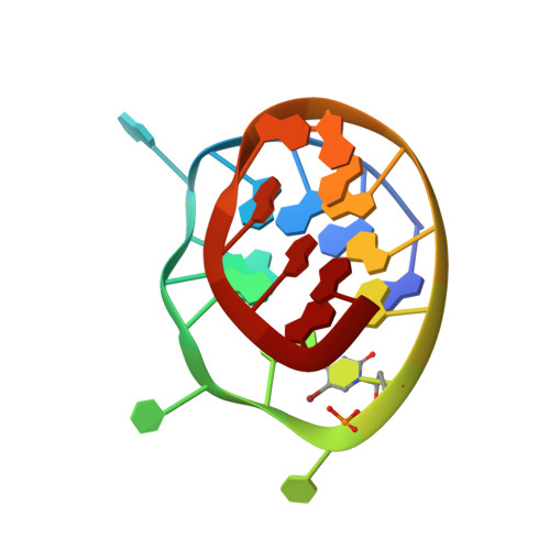
Crystal structure of a c-kit promoter quadruplex reveals the structural role of metal ions and water molecules in maintaining loop conformation.

Wei, D.Parkinson, G.N.Reszka, A.P.Neidle, S.

(2012) Nucleic Acids Res 40: 4691-4700

  • DOI: https://doi.org/10.1093/nar/gks023
  • Primary Citation of Related Structures:  
    3QXR

  • PubMed Abstract: 

    We report here the 1.62 Å crystal structure of an intramolecular quadruplex DNA formed from a sequence in the promoter region of the c-kit gene. This is the first reported crystal structure of a promoter quadruplex and the first observation of localized magnesium ions in a quadruplex structure. The structure reveals that potassium and magnesium ions have an unexpected yet significant structural role in stabilizing particular quadruplex loops and grooves that is distinct from but in addition to the role of potassium ions in the ion channel at the centre of all quadruplex structures. The analysis also shows how ions cluster together with structured water molecules to stabilize the quadruplex arrangement. This particular quadruplex has been previously studied by NMR methods, and the present X-ray structure is in accord with the earlier topology assignment. However, as well as the observations of potassium and magnesium ions, the crystal structure has revealed a highly significant difference in the dimensions of the large cleft in the structure, which is a plausible target for small molecules. This difference can be understood by the stabilizing role of structured water networks.


  • Organizational Affiliation

    CRUK Biomolecular Structure Group, UCL School of Pharmacy, University College London, 29-39 Brunswick Square, WC1N 1AX, London, UK.


Macromolecules

Find similar nucleic acids by:  Sequence   |   3D Structure  

Entity ID: 1
MoleculeChains LengthOrganismImage
5'-D(*AP*GP*GP*GP*AP*GP*GP*GP*CP*GP*CP*(BRU)P*GP*GP*GP*AP*GP*GP*AP*GP*GP*G)-3'
A, B
22Homo sapiens
Sequence Annotations
Expand
  • Reference Sequence
Small Molecules
Ligands 2 Unique
IDChains Name / Formula / InChI Key2D Diagram3D Interactions
K
Query on K

Download Ideal Coordinates CCD File 
C [auth A]
D [auth A]
E [auth A]
F [auth A]
G [auth A]
C [auth A],
D [auth A],
E [auth A],
F [auth A],
G [auth A],
J [auth B],
K [auth B],
L [auth B],
M [auth B]
POTASSIUM ION
K
NPYPAHLBTDXSSS-UHFFFAOYSA-N
MG
Query on MG

Download Ideal Coordinates CCD File 
H [auth A],
I [auth A]
MAGNESIUM ION
Mg
JLVVSXFLKOJNIY-UHFFFAOYSA-N
Experimental Data & Validation

Experimental Data

  • Method: X-RAY DIFFRACTION
  • Resolution: 1.62 Å
  • R-Value Free: 0.213 
  • R-Value Work: 0.166 
  • R-Value Observed: 0.168 
  • Space Group: P 21 21 2
Unit Cell:
Length ( Å )Angle ( ˚ )
a = 37.57α = 90
b = 51.35β = 90
c = 58.34γ = 90
Software Package:
Software NamePurpose
DNAdata collection
PHASESphasing
REFMACrefinement
PDB_EXTRACTdata extraction
xia2data reduction
SCALAdata scaling

Structure Validation

View Full Validation Report


Entry History 

Deposition Data

Revision History  (Full details and data files)

  • Version 1.0: 2012-01-18
    Type: Initial release
  • Version 1.1: 2012-01-25
    Changes: Database references
  • Version 1.2: 2012-02-15
    Changes: Database references
  • Version 1.3: 2012-06-06
    Changes: Database references
  • Version 1.4: 2017-11-08
    Changes: Refinement description
  • Version 1.5: 2023-09-13
    Changes: Data collection, Database references, Derived calculations, Refinement description