3QVP

Crystal structure of glucose oxidase for space group C2221 at 1.2 A resolution


Experimental Data Snapshot

  • Method: X-RAY DIFFRACTION
  • Resolution: 1.20 Å
  • R-Value Free: 0.188 
  • R-Value Work: 0.178 
  • R-Value Observed: 0.178 

wwPDB Validation   3D Report Full Report


Ligand Structure Quality Assessment 


This is version 2.1 of the entry. See complete history


Literature

Probing oxygen activation sites in two flavoprotein oxidases using chloride as an oxygen surrogate.

Kommoju, P.R.Chen, Z.W.Bruckner, R.C.Mathews, F.S.Jorns, M.S.

(2011) Biochemistry 50: 5521-5534

  • DOI: https://doi.org/10.1021/bi200388g
  • Primary Citation of Related Structures:  
    3QSE, 3QSM, 3QSS, 3QVP, 3QVR

  • PubMed Abstract: 

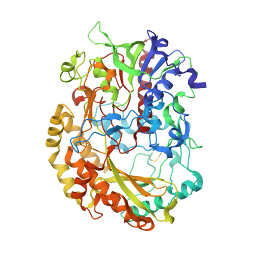
    A single basic residue above the si-face of the flavin ring is the site of oxygen activation in glucose oxidase (GOX) (His516) and monomeric sarcosine oxidase (MSOX) (Lys265). Crystal structures of both flavoenzymes exhibit a small pocket at the oxygen activation site that might provide a preorganized binding site for superoxide anion, an obligatory intermediate in the two-electron reduction of oxygen. Chloride binds at these polar oxygen activation sites, as judged by solution and structural studies. First, chloride forms spectrally detectable complexes with GOX and MSOX. The protonated form of His516 is required for tight binding of chloride to oxidized GOX and for rapid reaction of reduced GOX with oxygen. Formation of a binary MSOX·chloride complex requires Lys265 and is not observed with Lys265Met. Binding of chloride to MSOX does not affect the binding of a sarcosine analogue (MTA, methylthioactetate) above the re-face of the flavin ring. Definitive evidence is provided by crystal structures determined for a binary MSOX·chloride complex and a ternary MSOX·chloride·MTA complex. Chloride binds in the small pocket at a position otherwise occupied by a water molecule and forms hydrogen bonds to four ligands that are arranged in approximate tetrahedral geometry: Lys265:NZ, Arg49:NH1, and two water molecules, one of which is hydrogen bonded to FAD:N5. The results show that chloride (i) acts as an oxygen surrogate, (ii) is an effective probe of polar oxygen activation sites, and (iii) provides a valuable complementary tool to the xenon gas method that is used to map nonpolar oxygen-binding cavities.


  • Organizational Affiliation

    Department of Biochemistry and Molecular Biology, Drexel University College of Medicine, Philadelphia, Pennsylvania 19102, USA.


Macromolecules
Find similar proteins by:  (by identity cutoff)  |  3D Structure
Entity ID: 1
MoleculeChains Sequence LengthOrganismDetailsImage
Glucose oxidase583Aspergillus nigerMutation(s): 0 
Gene Names: gox
EC: 1.1.3.4
UniProt
Find proteins for P13006 (Aspergillus niger)
Explore P13006 
Go to UniProtKB:  P13006
Entity Groups  
Sequence Clusters30% Identity50% Identity70% Identity90% Identity95% Identity100% Identity
UniProt GroupP13006
Sequence Annotations
Expand
  • Reference Sequence
Oligosaccharides

Help

Entity ID: 2
MoleculeChains Length2D Diagram Glycosylation3D Interactions
alpha-D-mannopyranose-(1-2)-alpha-D-mannopyranose-(1-3)-beta-D-mannopyranose-(1-4)-2-acetamido-2-deoxy-beta-D-glucopyranose-(1-4)-2-acetamido-2-deoxy-beta-D-glucopyranose
B
5N-Glycosylation
Glycosylation Resources
GlyTouCan:  G42227JK
GlyCosmos:  G42227JK
GlyGen:  G42227JK
Small Molecules
Ligands 5 Unique
IDChains Name / Formula / InChI Key2D Diagram3D Interactions
FAD
Query on FAD

Download Ideal Coordinates CCD File 
F [auth A]FLAVIN-ADENINE DINUCLEOTIDE
C27 H33 N9 O15 P2
VWWQXMAJTJZDQX-UYBVJOGSSA-N
NAG
Query on NAG

Download Ideal Coordinates CCD File 
C [auth A],
D [auth A],
E [auth A]
2-acetamido-2-deoxy-beta-D-glucopyranose
C8 H15 N O6
OVRNDRQMDRJTHS-FMDGEEDCSA-N
PEG
Query on PEG

Download Ideal Coordinates CCD File 
H [auth A]DI(HYDROXYETHYL)ETHER
C4 H10 O3
MTHSVFCYNBDYFN-UHFFFAOYSA-N
GOL
Query on GOL

Download Ideal Coordinates CCD File 
G [auth A]GLYCEROL
C3 H8 O3
PEDCQBHIVMGVHV-UHFFFAOYSA-N
CL
Query on CL

Download Ideal Coordinates CCD File 
I [auth A]CHLORIDE ION
Cl
VEXZGXHMUGYJMC-UHFFFAOYSA-M
Experimental Data & Validation

Experimental Data

  • Method: X-RAY DIFFRACTION
  • Resolution: 1.20 Å
  • R-Value Free: 0.188 
  • R-Value Work: 0.178 
  • R-Value Observed: 0.178 
  • Space Group: C 2 2 21
Unit Cell:
Length ( Å )Angle ( ˚ )
a = 69.56α = 90
b = 119.147β = 90
c = 142.188γ = 90
Software Package:
Software NamePurpose
ADSCdata collection
MOLREPphasing
CNSrefinement
HKL-2000data reduction
HKL-2000data scaling

Structure Validation

View Full Validation Report


Ligand Structure Quality Assessment 


Entry History 

Deposition Data

Revision History  (Full details and data files)

  • Version 1.0: 2011-06-01
    Type: Initial release
  • Version 1.1: 2011-07-13
    Changes: Version format compliance
  • Version 2.0: 2020-07-29
    Type: Remediation
    Reason: Carbohydrate remediation
    Changes: Atomic model, Data collection, Derived calculations, Structure summary
  • Version 2.1: 2023-09-13
    Changes: Data collection, Database references, Refinement description, Structure summary