


Crystal Structure Analysis of Entamoeba histolytica Methyltransferase
Schulz, E.C., Roth, H.M., Ankri, S., Ficner, R.To be published.
Experimental Data Snapshot
Entity ID: 1 | |||||
|---|---|---|---|---|---|
| Molecule | Chains | Sequence Length | Organism | Details | Image |
| 5-cytosine DNA methyltransferase | 327 | Entamoeba histolytica | Mutation(s): 0 Gene Names: meth, EHI_069140 EC: 2.1.1.37 | 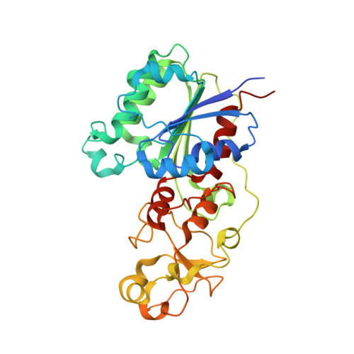 | |
UniProt | |||||
Find proteins for Q6Q386 (Entamoeba histolytica) Explore Q6Q386 Go to UniProtKB: Q6Q386 | |||||
Entity Groups | |||||
| Sequence Clusters | 30% Identity50% Identity70% Identity90% Identity95% Identity100% Identity | ||||
| UniProt Group | Q6Q386 | ||||
Sequence AnnotationsExpand | |||||
| |||||
| Ligands 2 Unique | |||||
|---|---|---|---|---|---|
| ID | Chains | Name / Formula / InChI Key | 2D Diagram | 3D Interactions | |
| SAH Query on SAH | B [auth A] | S-ADENOSYL-L-HOMOCYSTEINE C14 H20 N6 O5 S ZJUKTBDSGOFHSH-WFMPWKQPSA-N |  | ||
| SO4 Query on SO4 | C [auth A], D [auth A], E [auth A] | SULFATE ION O4 S QAOWNCQODCNURD-UHFFFAOYSA-L |  | ||
| Length ( Å ) | Angle ( ˚ ) |
|---|---|
| a = 46.97 | α = 90 |
| b = 46.97 | β = 90 |
| c = 303.24 | γ = 90 |
| Software Name | Purpose |
|---|---|
| XSCALE | data scaling |
| PHASER | phasing |
| PHENIX | refinement |
| PDB_EXTRACT | data extraction |
| DNA | data collection |
| XDS | data reduction |
RCSB PDB (citation) is hosted by
RCSB PDB is a member of the

