Significant stabilization of ribonuclease A by additive effects
Arnold, U., Schopfel, M.(2012) FEBS J 279: 2508-2519
- PubMed: 22594773
- DOI: https://doi.org/10.1111/j.1742-4658.2012.08632.x
- Primary Citation of Related Structures:
3QL1, 3QL2 - PubMed Abstract:
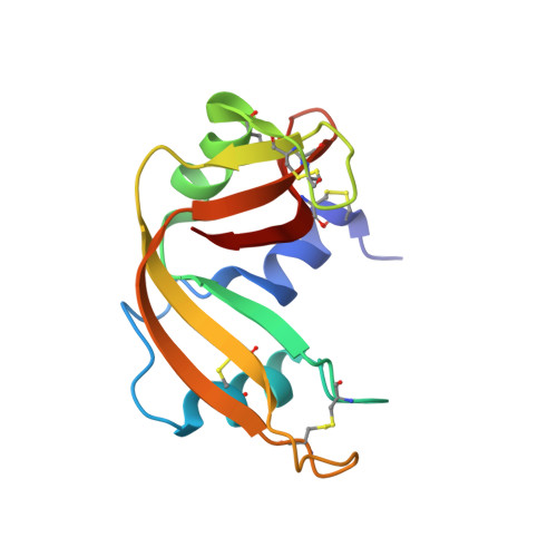
Among the strategies that employ genetic engineering to stabilize proteins, the introduction of disulfide bonds has proven to be a very potential approach. As, however, the replacement of amino acid residues by cysteines and the subsequent formation of the covalent bond can result in a severe deformation of the parental protein structure, the stabilization effect is strongly context dependent. Alternatively, the introduction of charged amino acid residues at the surface, which may result in the formation of extra ionic interactions or hydrogen bonds, provide propitious means for protein stabilization. The generation of an extra disulfide bond between residues 4 and 118 in ribonuclease A had resulted in a stabilization by 6 °C or 7 kJ mol(-1), which was mainly caused by a deceleration of the unfolding reaction [Pecher, P. & Arnold, U. (2009) Biophys Chem, 141, 21-28]. Here, Asp83 was replaced by Glu resulting in a comparable stabilization. Moreover, combination of both mutations led to an additive effect and the resulting ribonuclease A variant (T(m) ~ 76 °C, ΔG° ~ 53 kJ mol(-1)) is the most stable ribonuclease A variant described so far. The analysis of the crystal structure of A4C/D83E/V118C-ribonuclease A reveals the formation of a salt bridge between the γ-carboxyl group of Glu83 and the ε-amino group of Lys104.
Organizational Affiliation:
Institute of Biochemistry and Biotechnology, Martin-Luther University Halle-Wittenberg, Halle, Germany. ulrich.arnold@biochemtech.uni-halle.de