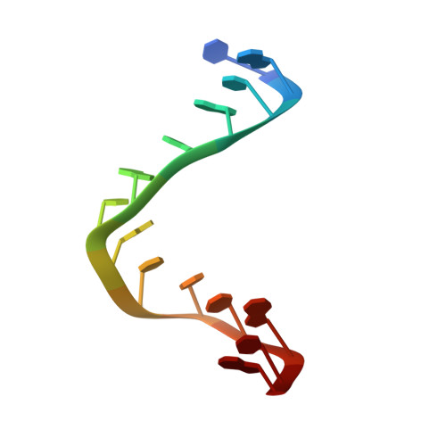
The structure of a full turn of an A-DNA duplex d(CGCGGGTACCCGCG)(2)
Venkadesh, S., Mandal, P.K., Gautham, N.(2011) Biochem Biophys Res Commun 407: 307-312
- PubMed: 21397589
- DOI: https://doi.org/10.1016/j.bbrc.2011.03.007
- Primary Citation of Related Structures:
3QK4 - PubMed Abstract:
We report the 2.6Å resolution crystal structure of the tetra-decamer d(CGCGGGTACCCGCG) in the tetragonal space group P4₃. This sequence contains the KpnI restriction site GGTACC in the centre which is flanked by alternating 'CG' sequences, and has a 'TA' step at the centre. These are features could favour the left-handed Z type helix. Despite this, overall the molecule has the A form. This is the first tetra-decamer crystallized in the A-DNA conformation, i.e. more than one full turn of the A helix. The crystallographic asymmetric unit consists of one tetra-decamer duplex. The helical twist and slide, as well as the base pair-base pair stacking interactions show alternations at the alternating pyrimidine-purine and purine-pyrimidine base steps. This variation is reminiscent of the dinucleotide repeat in left-handed Z-DNA helices. The crystal packing is unlike other A-DNA crystal structures, with each helix having a large number of contacts of many different types with symmetry-related neighbours.
Organizational Affiliation:
CAS in Crystallography and Biophysics, University of Madras, Chennai 600 025, Tamil Nadu, India.