3PM3

Bovine trypsin variant X(tripleIle227) in complex with small molecule inhibitor


Experimental Data Snapshot

  • Method: X-RAY DIFFRACTION
  • Resolution: 1.53 Å
  • R-Value Free: 0.181 
  • R-Value Work: 0.154 
  • R-Value Observed: 0.154 

wwPDB Validation   3D Report Full Report


Ligand Structure Quality Assessment 


This is version 1.5 of the entry. See complete history


Literature

Correlating structure and ligand affinity in drug discovery: a cautionary tale involving second shell residues.

Tziridis, A.Rauh, D.Neumann, P.Kolenko, P.Menzel, A.Brauer, U.Ursel, C.Steinmetzer, P.Sturzebecher, J.Schweinitz, A.Steinmetzer, T.Stubbs, M.T.

(2014) Biol Chem 395: 891-903

  • DOI: https://doi.org/10.1515/hsz-2014-0158
  • Primary Citation of Related Structures:  
    3PLB, 3PLK, 3PLP, 3PM3, 3PMJ, 3PWB, 3PWC, 3PYH, 3Q00, 3UNQ, 3UNS, 3UOP, 3UPE, 3UQO, 3UQV, 3UUZ, 3UWI, 3UY9, 3V12, 3V13

  • PubMed Abstract: 

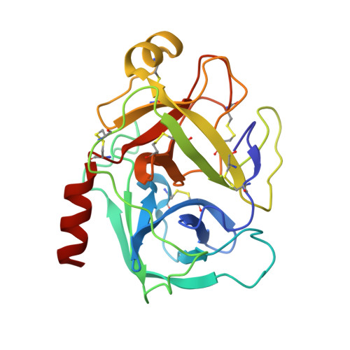
    A high-resolution crystallographic structure determination of a protein-ligand complex is generally accepted as the 'gold standard' for structure-based drug design, yet the relationship between structure and affinity is neither obvious nor straightforward. Here we analyze the interactions of a series of serine proteinase inhibitors with trypsin variants onto which the ligand-binding site of factor Xa has been grafted. Despite conservative mutations of only two residues not immediately in contact with ligands (second shell residues), significant differences in the affinity profiles of the variants are observed. Structural analyses demonstrate that these are due to multiple effects, including differences in the structure of the binding site, differences in target flexibility and differences in inhibitor binding modes. The data presented here highlight the myriad competing microscopic processes that contribute to protein-ligand interactions and emphasize the difficulties in predicting affinity from structure.


Macromolecules
Find similar proteins by:  (by identity cutoff)  |  3D Structure
Entity ID: 1
MoleculeChains Sequence LengthOrganismDetailsImage
Cationic trypsin223Bos taurusMutation(s): 8 
EC: 3.4.21.4
UniProt
Find proteins for P00760 (Bos taurus)
Explore P00760 
Go to UniProtKB:  P00760
Entity Groups  
Sequence Clusters30% Identity50% Identity70% Identity90% Identity95% Identity100% Identity
UniProt GroupP00760
Sequence Annotations
Expand
  • Reference Sequence
Small Molecules
Ligands 4 Unique
IDChains Name / Formula / InChI Key2D Diagram3D Interactions
TFN
Query on TFN

Download Ideal Coordinates CCD File 
C [auth A]N-(benzylsulfonyl)-O-tert-butyl-D-seryl-N-(4-carbamimidoylbenzyl)glycinamide
C24 H33 N5 O5 S
NAEBSNBHOZLKPM-HXUWFJFHSA-N
SO4
Query on SO4

Download Ideal Coordinates CCD File 
D [auth A],
G [auth A],
H [auth A]
SULFATE ION
O4 S
QAOWNCQODCNURD-UHFFFAOYSA-L
GOL
Query on GOL

Download Ideal Coordinates CCD File 
E [auth A],
F [auth A]
GLYCEROL
C3 H8 O3
PEDCQBHIVMGVHV-UHFFFAOYSA-N
CA
Query on CA

Download Ideal Coordinates CCD File 
B [auth A]CALCIUM ION
Ca
BHPQYMZQTOCNFJ-UHFFFAOYSA-N
Biologically Interesting Molecules (External Reference) 1 Unique
Experimental Data & Validation

Experimental Data

  • Method: X-RAY DIFFRACTION
  • Resolution: 1.53 Å
  • R-Value Free: 0.181 
  • R-Value Work: 0.154 
  • R-Value Observed: 0.154 
  • Space Group: P 31 2 1
Unit Cell:
Length ( Å )Angle ( ˚ )
a = 54.502α = 90
b = 54.502β = 90
c = 111.708γ = 120
Software Package:
Software NamePurpose
MxCuBEdata collection
AMoREphasing
REFMACrefinement
HKL-2000data reduction
SCALEPACKdata scaling

Structure Validation

View Full Validation Report


Ligand Structure Quality Assessment 


Entry History 

Deposition Data

Revision History  (Full details and data files)

  • Version 1.0: 2011-12-07
    Type: Initial release
  • Version 1.1: 2012-01-18
    Changes: Non-polymer description
  • Version 1.2: 2012-12-12
    Changes: Other
  • Version 1.3: 2014-07-16
    Changes: Database references
  • Version 1.4: 2014-09-24
    Changes: Database references
  • Version 1.5: 2023-09-06
    Changes: Data collection, Database references, Derived calculations, Refinement description