3PIJ

beta-fructofuranosidase from Bifidobacterium longum - complex with fructose


Experimental Data Snapshot

  • Method: X-RAY DIFFRACTION
  • Resolution: 1.80 Å
  • R-Value Free: 0.208 
  • R-Value Work: 0.153 
  • R-Value Observed: 0.154 

wwPDB Validation   3D Report Full Report


Ligand Structure Quality Assessment 


This is version 1.3 of the entry. See complete history


Literature

Crystal structures of the apo form of beta-fructofuranosidase from Bifidobacterium longum and its complex with fructose

Bujacz, A.Jedrzejczak-Krzepkowska, M.Bielecki, S.Redzynia, I.Bujacz, G.

(2011) FEBS J 278: 1728-1744

  • DOI: https://doi.org/10.1111/j.1742-4658.2011.08098.x
  • Primary Citation of Related Structures:  
    3PIG, 3PIJ

  • PubMed Abstract: 

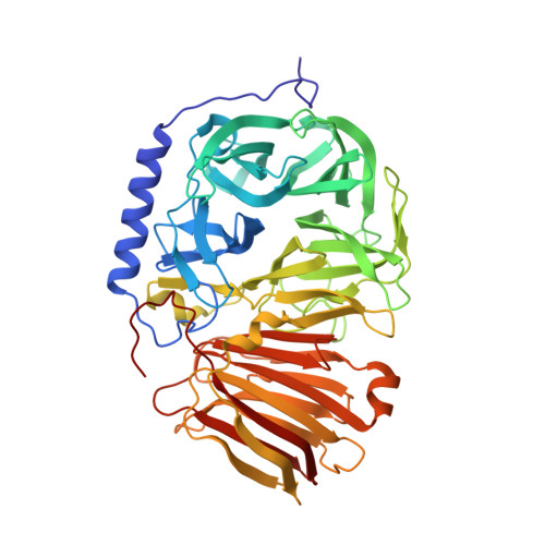
    We solved the 1.8 Å crystal structure of β-fructofuranosidase from Bifidobacterium longum KN29.1 - a unique enzyme that allows these probiotic bacteria to function in the human digestive system. The sequence of β-fructofuranosidase classifies it as belonging to the glycoside hydrolase family 32 (GH32). GH32 enzymes show a wide range of substrate specificity and different functions in various organisms. All enzymes from this family share a similar fold, containing two domains: an N-terminal five-bladed β-propeller and a C-terminal β-sandwich module. The active site is located in the centre of the β-propeller domain, in the bottom of a 'funnel'. The binding site, -1, responsible for tight fructose binding, is highly conserved among the GH32 enzymes. Bifidobacterium longum KN29.1 β-fructofuranosidase has a 35-residue elongation of the N-terminus containing a five-turn α-helix, which distinguishes it from the other known members of the GH32 family. This new structural element could be one of the functional modifications of the enzyme that allows the bacteria to act in a human digestive system. We also solved the 1.8 Å crystal structure of the β-fructofuranosidase complex with β-D-fructose, a hydrolysis product obtained by soaking apo crystal in raffinose.


  • Organizational Affiliation

    Institute of Technical Biochemistry, Faculty of Biotechnology and Food Sciences, Technical University of Lodz, Lodz, Poland. anna.bujacz@p.lodz.pl


Macromolecules
Find similar proteins by:  (by identity cutoff)  |  3D Structure
Entity ID: 1
MoleculeChains Sequence LengthOrganismDetailsImage
Beta-fructofuranosidase
A, B
526Bifidobacterium longumMutation(s): 1 
Gene Names: EF216677
EC: 3.2.1.26
UniProt
Find proteins for A2TLS9 (Bifidobacterium longum)
Explore A2TLS9 
Go to UniProtKB:  A2TLS9
Entity Groups  
Sequence Clusters30% Identity50% Identity70% Identity90% Identity95% Identity100% Identity
UniProt GroupA2TLS9
Sequence Annotations
Expand
  • Reference Sequence
Experimental Data & Validation

Experimental Data

  • Method: X-RAY DIFFRACTION
  • Resolution: 1.80 Å
  • R-Value Free: 0.208 
  • R-Value Work: 0.153 
  • R-Value Observed: 0.154 
  • Space Group: P 31 2 1
Unit Cell:
Length ( Å )Angle ( ˚ )
a = 86.765α = 90
b = 86.765β = 90
c = 223.868γ = 120
Software Package:
Software NamePurpose
HKL-2000data collection
REFMACrefinement
HKL-2000data reduction
HKL-2000data scaling
REFMACphasing

Structure Validation

View Full Validation Report


Ligand Structure Quality Assessment 


Entry History 

Deposition Data

Revision History  (Full details and data files)

  • Version 1.0: 2011-04-27
    Type: Initial release
  • Version 1.1: 2011-07-13
    Changes: Version format compliance
  • Version 1.2: 2020-07-29
    Type: Remediation
    Reason: Carbohydrate remediation
    Changes: Data collection, Database references, Derived calculations, Structure summary
  • Version 1.3: 2023-09-06
    Changes: Data collection, Database references, Refinement description, Structure summary